- PDB-3u8p: Cytochrome b562 integral fusion with EGFP -
+
Open data
ID or keywords:
Loading...
-
Basic information
Entry
Database: PDB / ID: 3u8p
Title
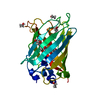
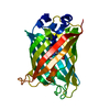
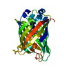
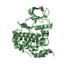
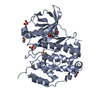
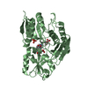
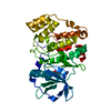
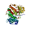
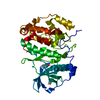
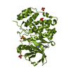
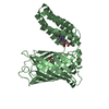
Cytochrome b562 integral fusion with EGFP
Components
Cytochrome b562 integral fusion with enhanced green fluorescent protein
Keywords
FLUORESCENT PROTEIN / ELECTRON TRANSPORT / directed evolution / domain insertion / energy transfer / fluorescence quenching
Function / homology
Function and homology information
bioluminescence / generation of precursor metabolites and energy / electron transport chain / electron transfer activity / periplasmic space / iron ion binding / heme binding Similarity search - Function
Cytochrome c/b562 / Green Fluorescent Protein / Green fluorescent protein / Cytochrome b562 / Cytochrome b562 / Cytochrome c/b562 / Green fluorescent protein, GFP / Green fluorescent protein-related / Green fluorescent protein / Green fluorescent protein ...Cytochrome c/b562 / Green Fluorescent Protein / Green fluorescent protein / Cytochrome b562 / Cytochrome b562 / Cytochrome c/b562 / Green fluorescent protein, GFP / Green fluorescent protein-related / Green fluorescent protein / Green fluorescent protein / Four Helix Bundle (Hemerythrin (Met), subunit A) / Up-down Bundle / Beta Barrel / Mainly Beta / Mainly Alpha Similarity search - Domain/homology
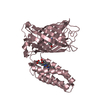
A: Cytochrome b562 integral fusion with enhanced green fluorescent protein B: Cytochrome b562 integral fusion with enhanced green fluorescent protein C: Cytochrome b562 integral fusion with enhanced green fluorescent protein hetero molecules
Mass: 18.015 Da / Num. of mol.: 344 / Source method: isolated from a natural source / Formula: H2O
Has protein modification
Y
Sequence details
RESIDUE 65 SER OF THE GREEN FLUORESCENT PROTEIN WAS MUTATED TO THR. RESIDUES 65 THR, 66 TYR, 67 GLY ...RESIDUE 65 SER OF THE GREEN FLUORESCENT PROTEIN WAS MUTATED TO THR. RESIDUES 65 THR, 66 TYR, 67 GLY FORMED A CHROMOPHORE CRO.
-
Experimental details
-
Experiment
Experiment
Method: X-RAY DIFFRACTION / Number of used crystals: 1
-
Sample preparation
Crystal
Density Matthews: 3.09 Å3/Da / Density % sol: 60.24 %
Crystal grow
Method: vapor diffusion / pH: 6.4 Details: 0.1 M MES/NAOH, PH 6.4, 200 MM magnesium acetate and 20% (W/V) PEG 8000; for cryoprotection 16% glycerol was added to the reservoir buffer, VAPOR DIFFUSION
Resolution: 2.75→28.94 Å / Cor.coef. Fo:Fc: 0.946 / Cor.coef. Fo:Fc free: 0.922 / Cross valid method: THROUGHOUT / ESU R Free: 0.334 / Stereochemistry target values: MAXIMUM LIKELIHOOD Details: TLS REFINEMENT HAS BEEN USED. HYDROGENS HAVE BEEN ADDED IN THE RIDING POSITIONS. THE RESOLUTION OF THIS STRUCTURE IS INSUFFICIENT TO JUDGE THE DIHEDRAL ANGLES OF THE HEME VINYLS. WE WOULD ...Details: TLS REFINEMENT HAS BEEN USED. HYDROGENS HAVE BEEN ADDED IN THE RIDING POSITIONS. THE RESOLUTION OF THIS STRUCTURE IS INSUFFICIENT TO JUDGE THE DIHEDRAL ANGLES OF THE HEME VINYLS. WE WOULD HAVE EXPECTED THAT THE VINYLS LIE IN THE PLANE OF THE PORPHYRIN RING, BUT NOTICED THAT THIS IS NOT THE CASE IN SEVERAL HIGH RESOLUTION HEME STRUCTURES (SEE ALSO REID ET AL., J. AM. CHEM. SOC. 1986, 108, 8197-8201) AND HAVE THEREFORE NOT RESTRAINED THE VINYL DIHEDRAL ANGLES.
Rfactor
Num. reflection
% reflection
Selection details
Rfree
0.24192
1882
5.1 %
RANDOM
Rwork
0.20099
-
-
-
obs
0.20317
34888
99.35 %
-
Solvent computation
Ion probe radii: 0.8 Å / Shrinkage radii: 0.8 Å / VDW probe radii: 1.4 Å / Solvent model: MASK
In the structure databanks used in Yorodumi, some data are registered as the other names, "COVID-19 virus" and "2019-nCoV". Here are the details of the virus and the list of structure data.
Jan 31, 2019. EMDB accession codes are about to change! (news from PDBe EMDB page)
EMDB accession codes are about to change! (news from PDBe EMDB page)
The allocation of 4 digits for EMDB accession codes will soon come to an end. Whilst these codes will remain in use, new EMDB accession codes will include an additional digit and will expand incrementally as the available range of codes is exhausted. The current 4-digit format prefixed with “EMD-” (i.e. EMD-XXXX) will advance to a 5-digit format (i.e. EMD-XXXXX), and so on. It is currently estimated that the 4-digit codes will be depleted around Spring 2019, at which point the 5-digit format will come into force.
The EM Navigator/Yorodumi systems omit the EMD- prefix.
Related info.:Q: What is EMD? / ID/Accession-code notation in Yorodumi/EM Navigator
Yorodumi is a browser for structure data from EMDB, PDB, SASBDB, etc.
This page is also the successor to EM Navigator detail page, and also detail information page/front-end page for Omokage search.
The word "yorodu" (or yorozu) is an old Japanese word meaning "ten thousand". "mi" (miru) is to see.
Related info.:EMDB / PDB / SASBDB / Comparison of 3 databanks / Yorodumi Search / Aug 31, 2016. New EM Navigator & Yorodumi / Yorodumi Papers / Jmol/JSmol / Function and homology information / Changes in new EM Navigator and Yorodumi
 Movie
Movie Controller


 Open data
 Basic information
 Components
 Keywords
 Function and homology information
 Aequorea victoria (jellyfish)
 X-RAY DIFFRACTION /
  Authors
 Citation
 Structure visualization
 Downloads & links
 Other downloads
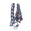

 PDBj
PDBj


 Assembly
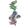


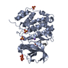
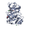
 Mass: 616.487 Da / Num. of mol.: 3 / Source method: obtained synthetically / Formula: C34H32FeN4O4
 Mass: 18.015 Da / Num. of mol.: 344 / Source method: isolated from a natural source / Formula: H2O
 Sample preparation
 / Beamline: I02 / Wavelength: 0.97949 / Wavelength: 0.97949 Å
 Processing