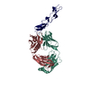
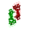
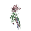
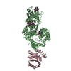
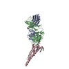
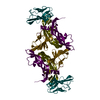
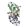
- PDB-3u22: Crystal structure of a putative HmuY_like heme binding protein (B... -
+
Open data
ID or keywords:
Loading...
-
Basic information
Entry
Database: PDB / ID: 3u22
Title
Crystal structure of a putative HmuY_like heme binding protein (BVU_2192) from Bacteroides vulgatus ATCC 8482 at 2.12 A resolution
Components
putative HmuY_like heme binding protein
Keywords
HEME BINDING PROTEIN / transport / Structural Genomics / Joint Center for Structural Genomics / JCSG / Protein Structure Initiative / PSI-BIOLOGY / HEME-BINDING PROTEIN
Function / homology
HmuY protein / HmuY protein / Unknown ligand / HmuY family protein
Function and homology information
Biological species
Bacteroides vulgatus (bacteria)
Method
X-RAY DIFFRACTION / SYNCHROTRON / MAD / Resolution: 2.12 Å
Mass: 18.015 Da / Num. of mol.: 275 / Source method: isolated from a natural source / Formula: H2O
-
Details
Has protein modification
Y
Sequence details
THIS CONSTRUCT WAS EXPRESSED WITH A PURIFICATION TAG MGSDKIHHHHHHENLYFQG. THE TAG WAS REMOVED WITH ...THIS CONSTRUCT WAS EXPRESSED WITH A PURIFICATION TAG MGSDKIHHHHHHENLYFQG. THE TAG WAS REMOVED WITH TEV PROTEASE LEAVING ONLY A GLYCINE (0) FOLLOWED BY RESIDUES 20-220 OF THE TARGET SEQUENCE.
-
Experimental details
-
Experiment
Experiment
Method: X-RAY DIFFRACTION / Number of used crystals: 1
-
Sample preparation
Crystal grow
Temperature: 293 K / Method: vapor diffusion, sitting drop Details: 0.1M bicine pH 9, 1.6M ammonium sulfate, NANODROP, VAPOR DIFFUSION, SITTING DROP, temperature 293K
Type: MARMOSAIC 325 mm CCD / Detector: CCD / Date: Jul 20, 2011 Details: Flat mirror (vertical focusing); single crystal Si(111) bent monochromator (ho rizontal focusing)
Radiation
Monochromator: single crystal Si(111) bent / Protocol: MAD / Monochromatic (M) / Laue (L): M / Scattering type: x-ray
Radiation wavelength
ID
Wavelength (Å)
Relative weight
1
0.91837
1
2
0.97922
1
3
0.97889
1
Reflection
Resolution: 2.12→78.487 Å / Num. all: 60126 / Num. obs: 60126 / % possible obs: 100 % / Redundancy: 5.7 % / Rsym value: 0.118 / Net I/σ(I): 9.6
Reflection shell
Rmerge(I) obs: 0.01 / Diffraction-ID: 1
Resolution (Å)
Redundancy (%)
Mean I/σ(I) obs
Num. measured all
Num. unique all
Rsym value
% possible all
2.12-2.23
5.8
0.7
49900
8674
1.019
100
2.23-2.37
5.8
0.8
47314
8224
0.672
100
2.37-2.53
5.8
1.5
44828
7774
0.476
100
2.53-2.74
5.8
1.6
41594
7210
0.317
100
2.74-3
5.8
3.9
38416
6658
0.183
100
3-3.35
5.8
6.1
34804
6039
0.109
100
3.35-3.87
5.7
8.6
30829
5362
0.067
100
3.87-4.74
5.7
9.4
26031
4556
0.05
100
4.74-6.7
5.6
12.2
20166
3574
0.046
100
6.7-78.487
5.3
12.8
10974
2055
0.039
99.9
-
Phasing
Phasing
Method: MAD
-
Processing
Software
Name
Version
Classification
NB
MolProbity
3beta29
modelbuilding
PDB_EXTRACT
3.1
dataextraction
SOLVE
phasing
SCALA
3.3.15
datascaling
REFMAC
5.6.0117
refinement
MOSFLM
datareduction
Refinement
Method to determine structure: MAD / Resolution: 2.12→78.487 Å / Cor.coef. Fo:Fc: 0.971 / Cor.coef. Fo:Fc free: 0.97 / Occupancy max: 1 / Occupancy min: 0.5 / SU B: 5.417 / SU ML: 0.071 / Cross valid method: THROUGHOUT / σ(F): 0 / ESU R Free: 0.09 Stereochemistry target values: MAXIMUM LIKELIHOOD WITH PHASES Details: 1. HYDROGENS HAVE BEEN ADDED IN THE RIDING POSITIONS. 2. ATOM RECORD CONTAINS SUM OF TLS AND RESIDUAL B FACTORS. ANISOU RECORD CONTAINS SUM OF TLS AND RESIDUAL U FACTORS. 3. WATERS WERE ...Details: 1. HYDROGENS HAVE BEEN ADDED IN THE RIDING POSITIONS. 2. ATOM RECORD CONTAINS SUM OF TLS AND RESIDUAL B FACTORS. ANISOU RECORD CONTAINS SUM OF TLS AND RESIDUAL U FACTORS. 3. WATERS WERE EXCLUDED FROM AUTOMATIC TLS ASSIGNMENT. 4. A MET-INHIBITION PROTOCOL WAS USED FOR SELENOMETHIONINE INCORPORATION DURING PROTEIN EXPRESSION. THE OCCUPANCY OF THE SE ATOMS IN THE MSE RESIDUES WAS REDUCED TO 0.75 FOR THE REDUCED SCATTERING POWER DUE TO PARTIAL S-MET INCORPORATION. 5. SULFATE ION (SO4) AND GLYCEROL (GOL) MOLECULES FROM THE CRYSTALLIZATION/CRYOPROTECTION SOLUTION ARE MODELED. 6. AN UNKNOWN LIGAND (UNL) HAS BEEN MODELED. 7. RESIDUE MSE172 FROM BOTH CHAINS ARE LOCATED IN THE SAME REGION AND THEREFORE IT IS DIFFICULT TO MODEL THEIR SIDECHAINS.
Rfactor
Num. reflection
% reflection
Selection details
Rfree
0.1724
3029
5 %
RANDOM
Rwork
0.1647
-
-
-
obs
0.1651
60066
99.87 %
-
Solvent computation
Ion probe radii: 0.8 Å / Shrinkage radii: 0.8 Å / VDW probe radii: 1.2 Å / Solvent model: BABINET MODEL WITH MASK
In the structure databanks used in Yorodumi, some data are registered as the other names, "COVID-19 virus" and "2019-nCoV". Here are the details of the virus and the list of structure data.
Jan 31, 2019. EMDB accession codes are about to change! (news from PDBe EMDB page)
EMDB accession codes are about to change! (news from PDBe EMDB page)
The allocation of 4 digits for EMDB accession codes will soon come to an end. Whilst these codes will remain in use, new EMDB accession codes will include an additional digit and will expand incrementally as the available range of codes is exhausted. The current 4-digit format prefixed with “EMD-” (i.e. EMD-XXXX) will advance to a 5-digit format (i.e. EMD-XXXXX), and so on. It is currently estimated that the 4-digit codes will be depleted around Spring 2019, at which point the 5-digit format will come into force.
The EM Navigator/Yorodumi systems omit the EMD- prefix.
Related info.:Q: What is EMD? / ID/Accession-code notation in Yorodumi/EM Navigator
Yorodumi is a browser for structure data from EMDB, PDB, SASBDB, etc.
This page is also the successor to EM Navigator detail page, and also detail information page/front-end page for Omokage search.
The word "yorodu" (or yorozu) is an old Japanese word meaning "ten thousand". "mi" (miru) is to see.
Related info.:EMDB / PDB / SASBDB / Comparison of 3 databanks / Yorodumi Search / Aug 31, 2016. New EM Navigator & Yorodumi / Yorodumi Papers / Jmol/JSmol / Function and homology information / Changes in new EM Navigator and Yorodumi
 Movie
Movie Controller

 Yorodumi
 Open data
 Basic information
 Components
 Keywords
 Function and homology information
 Bacteroides vulgatus (bacteria)
 X-RAY DIFFRACTION /
  Authors
 Citation
 Structure visualization
 Downloads & links
 Other downloads

 PDBj
PDBj

 Assembly
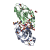


 Mass: 96.063 Da / Num. of mol.: 8 / Source method: obtained synthetically / Formula: SO4
 Mass: 92.094 Da / Num. of mol.: 8 / Source method: obtained synthetically / Formula: C3H8O3
 Mass: 35.453 Da / Num. of mol.: 1 / Source method: obtained synthetically / Formula: Cl
 Sample preparation
 / Beamline: BL11-1 / Wavelength: 0.91837,0.97922,0.97889
 Processing