 Movie
Movie Controller

[English] 日本語
 Yorodumi
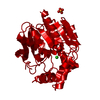
- PDB-3tzf: Crystal Structure of the Yersinia pestis Dihydropteroate Synthase... -
+ Open data

- Basic information
| Entry | Database: PDB / ID: 3tzf | ||||||
|---|---|---|---|---|---|---|---|
| Title | Crystal Structure of the Yersinia pestis Dihydropteroate Synthase with Sulfonamide Drug Complex. | ||||||
 Components Components | 7,8-dihydropteroate synthase | ||||||
 Keywords Keywords | TRANSFERASE/ANTIBIOTIC/INHIBITOR / Dihydropteroate synthase / sulfonamide complex / Tim barrel / TRANSFERASE-INHIBITOR-ANTIBIOTIC complex / TRANSFERASE-ANTIBIOTIC-INHIBITOR complex | ||||||
| Function / homology |  Function and homology information Function and homology informationdihydropteroate synthase / dihydropteroate synthase activity / folic acid biosynthetic process / tetrahydrofolate biosynthetic process / metal ion binding / cytosol Similarity search - Function | ||||||
| Biological species |   Yersinia pestis (bacteria) Yersinia pestis (bacteria) | ||||||
| Method |  X-RAY DIFFRACTION / SYNCHROTRON / MOLECULAR REPLACEMENT / molecular replacement / Resolution: 2.1 Å X-RAY DIFFRACTION / SYNCHROTRON / MOLECULAR REPLACEMENT / molecular replacement / Resolution: 2.1 Å | ||||||
 Authors Authors | Wu, Y. | ||||||
 Citation Citation | Journal: Science / Year: 2012 Title: Catalysis and sulfa drug resistance in dihydropteroate synthase. Authors: Yun, M.K. / Wu, Y. / Li, Z. / Zhao, Y. / Waddell, M.B. / Ferreira, A.M. / Lee, R.E. / Bashford, D. / White, S.W. | ||||||
| History |
|
- Structure visualization
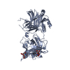
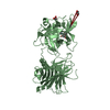
| Structure viewer | Molecule: MolmilJmol/JSmol |
|---|
- Downloads & links
-Download
| PDBx/mmCIF format | 3tzf.cif.gz | 222.3 KB | Display | PDBx/mmCIF format |
|---|---|---|---|---|
| PDB format | pdb3tzf.ent.gz | 178.2 KB | Display | PDB format |
| PDBx/mmJSON format | 3tzf.json.gz | Tree view | PDBx/mmJSON format | |
| Others |  Other downloads Other downloads |
-Validation report
| Arichive directory | https://data.pdbj.org/pub/pdb/validation_reports/tz/3tzfftp://data.pdbj.org/pub/pdb/validation_reports/tz/3tzf | HTTPS FTP |
|---|
-Related structure data
| Related structure data |  3tyaC  3tybC  3tycC  3tydC  3tyeC  3tyuC  3tyzC  3tznC  3v5oC C: citing same article ( |
|---|---|
| Similar structure data |


-Links
 PDBj
PDBj- Assembly
| Deposited unit | 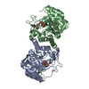
| ||||||||
|---|---|---|---|---|---|---|---|---|---|
| 1 |
| ||||||||
| Unit cell |
|
-Components
| #1: Protein | Mass: 30317.729 Da / Num. of mol.: 2 / Fragment: DHPS Source method: isolated from a genetically manipulated source Source: (gene. exp.) Yersinia pestis (bacteria) / Strain: KIM D27 / Gene: dhpS, folP, y0683, YPO3501, YP_0582 / Plasmid: pET28a / Production host: References: UniProt: Q7CKJ1, UniProt: A0A2S9PLG4*PLUS, dihydropteroate synthase #2: Chemical |   Mass: 353.123 Da / Num. of mol.: 2 / Source method: obtained synthetically / Formula: C7H9N5O8P2 Mass: 353.123 Da / Num. of mol.: 2 / Source method: obtained synthetically / Formula: C7H9N5O8P2#3: Chemical |   Mass: 24.305 Da / Num. of mol.: 2 / Source method: obtained synthetically / Formula: Mg Mass: 24.305 Da / Num. of mol.: 2 / Source method: obtained synthetically / Formula: Mg#4: Chemical | ChemComp-08D / |   Mass: 253.278 Da / Num. of mol.: 1 / Source method: obtained synthetically / Formula: C10H11N3O3S / Comment: antibiotic*YM Mass: 253.278 Da / Num. of mol.: 1 / Source method: obtained synthetically / Formula: C10H11N3O3S / Comment: antibiotic*YM#5: Water | ChemComp-HOH / |  Mass: 18.015 Da / Num. of mol.: 207 / Source method: isolated from a natural source / Formula: H2O Mass: 18.015 Da / Num. of mol.: 207 / Source method: isolated from a natural source / Formula: H2O |
|---|
-Experimental details
-Experiment
| Experiment | Method: X-RAY DIFFRACTION / Number of used crystals: 1 |
|---|
- Sample preparation
| Crystal | Density Matthews: 2.2 Å3/Da / Density % sol: 44.01 % |
|---|---|
| Crystal grow | Temperature: 291 K / Method: vapor diffusion, sitting drop Details: PEG 20,000, MES(pH6.5), pH 6-7, vapor diffusion, sitting drop, temperature 291K PH range: 6-7 |
-Data collection
| Diffraction | Mean temperature: 100 K | |||||||||||||||||||||||||||||||||||||||||||||||||||||||||||||||||||||||||||||||||||||||||||||||||||||||||||||||||||||||||||||||||||||||||||||||||||
|---|---|---|---|---|---|---|---|---|---|---|---|---|---|---|---|---|---|---|---|---|---|---|---|---|---|---|---|---|---|---|---|---|---|---|---|---|---|---|---|---|---|---|---|---|---|---|---|---|---|---|---|---|---|---|---|---|---|---|---|---|---|---|---|---|---|---|---|---|---|---|---|---|---|---|---|---|---|---|---|---|---|---|---|---|---|---|---|---|---|---|---|---|---|---|---|---|---|---|---|---|---|---|---|---|---|---|---|---|---|---|---|---|---|---|---|---|---|---|---|---|---|---|---|---|---|---|---|---|---|---|---|---|---|---|---|---|---|---|---|---|---|---|---|---|---|---|---|---|
| Diffraction source | Source: SYNCHROTRON / Site: APS  / Beamline: 22-BM / Wavelength: 0.97954 Å / Beamline: 22-BM / Wavelength: 0.97954 Å | |||||||||||||||||||||||||||||||||||||||||||||||||||||||||||||||||||||||||||||||||||||||||||||||||||||||||||||||||||||||||||||||||||||||||||||||||||
| Detector | Type: MARMOSAIC 225 mm CCD / Detector: CCD / Date: Apr 17, 2011 | |||||||||||||||||||||||||||||||||||||||||||||||||||||||||||||||||||||||||||||||||||||||||||||||||||||||||||||||||||||||||||||||||||||||||||||||||||
| Radiation | Monochromator: Si-220 / Protocol: SINGLE WAVELENGTH / Monochromatic (M) / Laue (L): M / Scattering type: x-ray | |||||||||||||||||||||||||||||||||||||||||||||||||||||||||||||||||||||||||||||||||||||||||||||||||||||||||||||||||||||||||||||||||||||||||||||||||||
| Radiation wavelength | Wavelength: 0.97954 Å / Relative weight: 1 | |||||||||||||||||||||||||||||||||||||||||||||||||||||||||||||||||||||||||||||||||||||||||||||||||||||||||||||||||||||||||||||||||||||||||||||||||||
| Reflection | Resolution: 2.1→50 Å / Num. obs: 30631 / % possible obs: 98.5 % / Observed criterion σ(I): -3 / Redundancy: 4.8 % / Rmerge(I) obs: 0.074 / Χ2: 0.849 / Net I/σ(I): 8.3 | |||||||||||||||||||||||||||||||||||||||||||||||||||||||||||||||||||||||||||||||||||||||||||||||||||||||||||||||||||||||||||||||||||||||||||||||||||
| Reflection shell |
|
-Phasing
| Phasing | Method: molecular replacement |
|---|
- Processing
| Software |
| |||||||||||||||||||||||||||||||||||||||||||||||||||||||||||||||||||||||||||||||||||||||||||||||||||||||||||||||||||||||||||||
|---|---|---|---|---|---|---|---|---|---|---|---|---|---|---|---|---|---|---|---|---|---|---|---|---|---|---|---|---|---|---|---|---|---|---|---|---|---|---|---|---|---|---|---|---|---|---|---|---|---|---|---|---|---|---|---|---|---|---|---|---|---|---|---|---|---|---|---|---|---|---|---|---|---|---|---|---|---|---|---|---|---|---|---|---|---|---|---|---|---|---|---|---|---|---|---|---|---|---|---|---|---|---|---|---|---|---|---|---|---|---|---|---|---|---|---|---|---|---|---|---|---|---|---|---|---|---|
| Refinement | Method to determine structure: MOLECULAR REPLACEMENT / Resolution: 2.1→50 Å / Cor.coef. Fo:Fc: 0.942 / Cor.coef. Fo:Fc free: 0.92 / Occupancy max: 1 / Occupancy min: 0.5 / SU B: 10.676 / SU ML: 0.13 / Cross valid method: THROUGHOUT / σ(F): 0 / ESU R Free: 0.201 / Stereochemistry target values: MAXIMUM LIKELIHOOD Details: HYDROGENS HAVE BEEN ADDED IN THE RIDING POSITIONS U VALUES : RESIDUAL ONLY
| |||||||||||||||||||||||||||||||||||||||||||||||||||||||||||||||||||||||||||||||||||||||||||||||||||||||||||||||||||||||||||||
| Solvent computation | Ion probe radii: 0.8 Å / Shrinkage radii: 0.8 Å / VDW probe radii: 1.4 Å / Solvent model: MASK | |||||||||||||||||||||||||||||||||||||||||||||||||||||||||||||||||||||||||||||||||||||||||||||||||||||||||||||||||||||||||||||
| Displacement parameters | Biso max: 68.9 Å2 / Biso mean: 33.2524 Å2 / Biso min: 7.7 Å2
| |||||||||||||||||||||||||||||||||||||||||||||||||||||||||||||||||||||||||||||||||||||||||||||||||||||||||||||||||||||||||||||
| Refinement step | Cycle: LAST / Resolution: 2.1→50 Å
| |||||||||||||||||||||||||||||||||||||||||||||||||||||||||||||||||||||||||||||||||||||||||||||||||||||||||||||||||||||||||||||
| Refine LS restraints |
| |||||||||||||||||||||||||||||||||||||||||||||||||||||||||||||||||||||||||||||||||||||||||||||||||||||||||||||||||||||||||||||
| LS refinement shell | Resolution: 2.1→2.152 Å / Total num. of bins used: 20
| |||||||||||||||||||||||||||||||||||||||||||||||||||||||||||||||||||||||||||||||||||||||||||||||||||||||||||||||||||||||||||||
| Refinement TLS params. | Method: refined / Refine-ID: X-RAY DIFFRACTION
| |||||||||||||||||||||||||||||||||||||||||||||||||||||||||||||||||||||||||||||||||||||||||||||||||||||||||||||||||||||||||||||
| Refinement TLS group |
|
