 Movie
Movie Controller


+ Open data

- Basic information
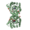
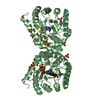
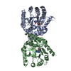
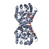
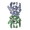
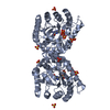
| Entry | Database: PDB / ID: 3tye | ||||||
|---|---|---|---|---|---|---|---|
| Title | Dihydropteroate Synthase in complex with DHP-STZ | ||||||
 Components Components | Dihydropteroate synthase | ||||||
 Keywords Keywords | TRANSFERASE / anthracis / folate biosynthesis / dihydropteroate / pterine / tim barrel | ||||||
| Function / homology |  Function and homology information Function and homology informationdihydropteroate synthase / dihydropteroate synthase activity / folic acid biosynthetic process / tetrahydrofolate biosynthetic process / metal ion binding / cytosol Similarity search - Function | ||||||
| Biological species |  | ||||||
| Method |  X-RAY DIFFRACTION / SYNCHROTRON / MOLECULAR REPLACEMENT / Resolution: 2.3 Å X-RAY DIFFRACTION / SYNCHROTRON / MOLECULAR REPLACEMENT / Resolution: 2.3 Å | ||||||
 Authors Authors | Yun, M.-K. / White, S.W. | ||||||
 Citation Citation | Journal: Science / Year: 2012 Title: Catalysis and sulfa drug resistance in dihydropteroate synthase. Authors: Yun, M.K. / Wu, Y. / Li, Z. / Zhao, Y. / Waddell, M.B. / Ferreira, A.M. / Lee, R.E. / Bashford, D. / White, S.W. | ||||||
| History |
|
- Structure visualization
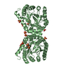
| Structure viewer | Molecule: MolmilJmol/JSmol |
|---|
- Downloads & links
-Download
| PDBx/mmCIF format | 3tye.cif.gz | 225.3 KB | Display | PDBx/mmCIF format |
|---|---|---|---|---|
| PDB format | pdb3tye.ent.gz | 183.6 KB | Display | PDB format |
| PDBx/mmJSON format | 3tye.json.gz | Tree view | PDBx/mmJSON format | |
| Others |  Other downloads Other downloads |
-Validation report
| Arichive directory | https://data.pdbj.org/pub/pdb/validation_reports/ty/3tyeftp://data.pdbj.org/pub/pdb/validation_reports/ty/3tye | HTTPS FTP |
|---|
-Related structure data
| Related structure data |  3tyaC  3tybC  3tycC  3tydC  3tyuC  3tyzC  3tzfC  3tznC  3v5oC  1twsS S: Starting model for refinement C: citing same article ( |
|---|---|
| Similar structure data |


-Links
 PDBj
PDBj
- Assembly
| Deposited unit | 
| ||||||||
|---|---|---|---|---|---|---|---|---|---|
| 1 | 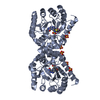
| ||||||||
| 2 | 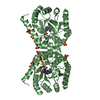
| ||||||||
| Unit cell |
|
-Components
-Protein , 1 types, 2 molecules AB
| #1: Protein | Mass: 32883.734 Da / Num. of mol.: 2 / Fragment: DIHYDROPTEROATE SYNTHASE Source method: isolated from a genetically manipulated source Source: (gene. exp.) |
|---|
-Non-polymers , 5 types, 96 molecules 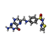


| #2: Chemical | ChemComp-XTZ /  Mass: 432.480 Da / Num. of mol.: 1 / Source method: obtained synthetically / Formula: C16H16N8O3S2 Mass: 432.480 Da / Num. of mol.: 1 / Source method: obtained synthetically / Formula: C16H16N8O3S2 | ||||||
|---|---|---|---|---|---|---|---|
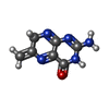
| #3: Chemical | ChemComp-SO4 /  Mass: 96.063 Da / Num. of mol.: 9 / Source method: obtained synthetically / Formula: SO4 Mass: 96.063 Da / Num. of mol.: 9 / Source method: obtained synthetically / Formula: SO4#4: Chemical | ChemComp-XHP / |  Mass: 177.163 Da / Num. of mol.: 1 / Source method: obtained synthetically / Formula: C7H7N5O Mass: 177.163 Da / Num. of mol.: 1 / Source method: obtained synthetically / Formula: C7H7N5O#5: Chemical | ChemComp-YTZ / |  Mass: 255.317 Da / Num. of mol.: 1 / Source method: obtained synthetically / Formula: C9H9N3O2S2 Mass: 255.317 Da / Num. of mol.: 1 / Source method: obtained synthetically / Formula: C9H9N3O2S2#6: Water | ChemComp-HOH / | Mass: 18.015 Da / Num. of mol.: 84 / Source method: isolated from a natural source / Formula: H2O |
-Experimental details
-Experiment
| Experiment | Method: X-RAY DIFFRACTION / Number of used crystals: 1 |
|---|
- Sample preparation
| Crystal | Density Matthews: 2.74 Å3/Da / Density % sol: 55.15 % |
|---|---|
| Crystal grow | Temperature: 291 K / Method: vapor diffusion, hanging drop / pH: 9 Details: LITHIUM SULFATE, Bis-Tris propane, pH 9.0, VAPOR DIFFUSION, HANGING DROP, temperature 291.0K |
-Data collection
| Diffraction | Mean temperature: 100 K |
|---|---|
| Diffraction source | Source: SYNCHROTRON / Site: APS  / Beamline: 22-ID / Wavelength: 1 Å / Beamline: 22-ID / Wavelength: 1 Å |
| Detector | Type: MARMOSAIC 300 mm CCD / Detector: CCD / Date: Dec 3, 2010 |
| Radiation | Protocol: SINGLE WAVELENGTH / Monochromatic (M) / Laue (L): M / Scattering type: x-ray |
| Radiation wavelength | Wavelength: 1 Å / Relative weight: 1 |
| Reflection | Resolution: 2.3→50 Å / Num. obs: 32382 / % possible obs: 95.2 % / Observed criterion σ(I): -3 / Redundancy: 9.2 % / Rsym value: 0.062 / Net I/σ(I): 30.5 |
| Reflection shell | Resolution: 2.3→2.38 Å / Redundancy: 8.9 % / Mean I/σ(I) obs: 5 / Num. unique all: 2520 / Rsym value: 0.219 / % possible all: 76 |
- Processing
| Software |
| |||||||||||||||||||||||||||||||||||||||||||||||||||||||||||||||||||||||||||
|---|---|---|---|---|---|---|---|---|---|---|---|---|---|---|---|---|---|---|---|---|---|---|---|---|---|---|---|---|---|---|---|---|---|---|---|---|---|---|---|---|---|---|---|---|---|---|---|---|---|---|---|---|---|---|---|---|---|---|---|---|---|---|---|---|---|---|---|---|---|---|---|---|---|---|---|---|
| Refinement | Method to determine structure: MOLECULAR REPLACEMENT Starting model: PDB ENTRY 1TWS Resolution: 2.3→45.66 Å / Cor.coef. Fo:Fc: 0.947 / Cor.coef. Fo:Fc free: 0.93 / Occupancy max: 1 / Occupancy min: 0.5 / SU B: 33.846 / SU ML: 0.325 / Cross valid method: THROUGHOUT / σ(F): 0 / ESU R: 0.341 / ESU R Free: 0.262 / Stereochemistry target values: MAXIMUM LIKELIHOOD
| |||||||||||||||||||||||||||||||||||||||||||||||||||||||||||||||||||||||||||
| Solvent computation | Ion probe radii: 0.8 Å / Shrinkage radii: 0.8 Å / VDW probe radii: 1.4 Å / Solvent model: MASK | |||||||||||||||||||||||||||||||||||||||||||||||||||||||||||||||||||||||||||
| Displacement parameters | Biso max: 166.69 Å2 / Biso mean: 78.2592 Å2 / Biso min: 2 Å2
| |||||||||||||||||||||||||||||||||||||||||||||||||||||||||||||||||||||||||||
| Refinement step | Cycle: LAST / Resolution: 2.3→45.66 Å
| |||||||||||||||||||||||||||||||||||||||||||||||||||||||||||||||||||||||||||
| Refine LS restraints |
| |||||||||||||||||||||||||||||||||||||||||||||||||||||||||||||||||||||||||||
| LS refinement shell | Resolution: 2.3→2.36 Å / Total num. of bins used: 20
| |||||||||||||||||||||||||||||||||||||||||||||||||||||||||||||||||||||||||||
| Refinement TLS params. | Method: refined / Refine-ID: X-RAY DIFFRACTION
| |||||||||||||||||||||||||||||||||||||||||||||||||||||||||||||||||||||||||||
| Refinement TLS group |
|
