 Movie
Movie Controller

[English] 日本語
 Yorodumi
- PDB-4utw: Structural characterisation of NanE, ManNac6P C2 epimerase, from ... -
+ Open data

- Basic information
| Entry | Database: PDB / ID: 4utw | ||||||
|---|---|---|---|---|---|---|---|
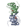
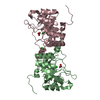
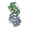
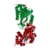
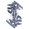
| Title | Structural characterisation of NanE, ManNac6P C2 epimerase, from Clostridium perfingens | ||||||
 Components Components | (PUTATIVE N-ACETYLMANNOSAMINE-6-PHOSPHATE 2-EPIMERASE) x 2 | ||||||
 Keywords Keywords | ISOMERASE / SUGAR 2-EPIMERASE / SIALIC ACID / SUGAR PHOSPHATE / ENZYME MECHANISM / CARBOHYDRATE / MUTAGENESIS / 1H NMR SPECTROSCOPY | ||||||
| Function / homology |  Function and homology information Function and homology informationN-acylglucosamine-6-phosphate 2-epimerase / N-acetylmannosamine catabolic process / N-acylglucosamine-6-phosphate 2-epimerase activity / N-acetylneuraminate catabolic process / carbohydrate metabolic process / cytosol Similarity search - Function | ||||||
| Biological species |  CLOSTRIDIUM PERFRINGENS STR. 13 (bacteria) CLOSTRIDIUM PERFRINGENS STR. 13 (bacteria) | ||||||
| Method |  X-RAY DIFFRACTION / SYNCHROTRON / MOLECULAR REPLACEMENT / Resolution: 1.9 Å X-RAY DIFFRACTION / SYNCHROTRON / MOLECULAR REPLACEMENT / Resolution: 1.9 Å | ||||||
 Authors Authors | Pelissier, M.C. / Sebban-Kreuzer, C. / Guerlesquin, F. / Brannigan, J.A. / Davies, G.J. / Bourne, Y. / Vincent, F. | ||||||
 Citation Citation | Journal: J.Biol.Chem. / Year: 2014 Title: Structural and Functional Characterization of the Clostridium Perfringens N-Acetylmannosamine-6-Phosphate 2-Epimerase Essential for the Sialic Acid Salvage Pathway Authors: Pelissier, M.C. / Sebban-Kreuzer, C. / Guerlesquin, F. / Brannigan, J.A. / Davies, G.J. / Bourne, Y. / Vincent, F. | ||||||
| History |
| ||||||
| Remark 700 | SHEET DETERMINATION METHOD: DSSP THE SHEETS PRESENTED AS "AA" IN EACH CHAIN ON SHEET RECORDS BELOW ... SHEET DETERMINATION METHOD: DSSP THE SHEETS PRESENTED AS "AA" IN EACH CHAIN ON SHEET RECORDS BELOW IS ACTUALLY AN 8-STRANDED BARREL THIS IS REPRESENTED BY A 9-STRANDED SHEET IN WHICH THE FIRST AND LAST STRANDS ARE IDENTICAL. THE SHEETS PRESENTED AS "BA" IN EACH CHAIN ON SHEET RECORDS BELOW IS ACTUALLY AN 8-STRANDED BARREL THIS IS REPRESENTED BY A 9-STRANDED SHEET IN WHICH THE FIRST AND LAST STRANDS ARE IDENTICAL. THE SHEETS PRESENTED AS "CA" IN EACH CHAIN ON SHEET RECORDS BELOW IS ACTUALLY AN 8-STRANDED BARREL THIS IS REPRESENTED BY A 9-STRANDED SHEET IN WHICH THE FIRST AND LAST STRANDS ARE IDENTICAL. THE SHEETS PRESENTED AS "DA" IN EACH CHAIN ON SHEET RECORDS BELOW IS ACTUALLY AN 8-STRANDED BARREL THIS IS REPRESENTED BY A 9-STRANDED SHEET IN WHICH THE FIRST AND LAST STRANDS ARE IDENTICAL. |
- Structure visualization
| Structure viewer | Molecule: MolmilJmol/JSmol |
|---|
- Downloads & links
-Download
| PDBx/mmCIF format | 4utw.cif.gz | 209.3 KB | Display | PDBx/mmCIF format |
|---|---|---|---|---|
| PDB format | pdb4utw.ent.gz | 168.3 KB | Display | PDB format |
| PDBx/mmJSON format | 4utw.json.gz | Tree view | PDBx/mmJSON format | |
| Others |  Other downloads Other downloads |
-Validation report
| Arichive directory | https://data.pdbj.org/pub/pdb/validation_reports/ut/4utwftp://data.pdbj.org/pub/pdb/validation_reports/ut/4utw | HTTPS FTP |
|---|
-Related structure data
| Related structure data | 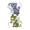 4uttSC  4utuC S: Starting model for refinement C: citing same article ( |
|---|---|
| Similar structure data |


-Links
 PDBj
PDBj
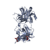
- Assembly
| Deposited unit | 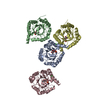
| |||||||||||||||||||||||||||||||||||||||||||||
|---|---|---|---|---|---|---|---|---|---|---|---|---|---|---|---|---|---|---|---|---|---|---|---|---|---|---|---|---|---|---|---|---|---|---|---|---|---|---|---|---|---|---|---|---|---|---|
| 1 | 
| |||||||||||||||||||||||||||||||||||||||||||||
| 2 | 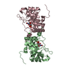
| |||||||||||||||||||||||||||||||||||||||||||||
| 3 | 
| |||||||||||||||||||||||||||||||||||||||||||||
| Unit cell |
| |||||||||||||||||||||||||||||||||||||||||||||
| Noncrystallographic symmetry (NCS) | NCS domain:
NCS domain segments:
|
-Components
| #1: Protein | Mass: 25164.900 Da / Num. of mol.: 2 Source method: isolated from a genetically manipulated source Details: 8 CHLORIDE IONS AND 4 ACTETATE MOLECULES ARE BOUND TO THE 4 MONOMERS Source: (gene. exp.) CLOSTRIDIUM PERFRINGENS STR. 13 (bacteria)Plasmid: PET-YSBLIC / Production host: References: UniProt: Q0TUP9, UniProt: Q8XNZ3*PLUS, N-acylglucosamine-6-phosphate 2-epimerase #2: Protein | Mass: 25113.832 Da / Num. of mol.: 2 Source method: isolated from a genetically manipulated source Details: 8 CHLORIDE IONS AND 4 ACTETATE MOLECULES ARE BOUND TO THE 4 MONOMERS Source: (gene. exp.) CLOSTRIDIUM PERFRINGENS STR. 13 (bacteria)Plasmid: PET-YSBLIC / Production host: References: UniProt: Q0TUP9, UniProt: Q8XNZ3*PLUS, N-acylglucosamine-6-phosphate 2-epimerase #3: Chemical | ChemComp-CL /   Mass: 35.453 Da / Num. of mol.: 4 / Source method: obtained synthetically / Formula: Cl Mass: 35.453 Da / Num. of mol.: 4 / Source method: obtained synthetically / Formula: Cl#4: Chemical | ChemComp-RFW /   Mass: 299.172 Da / Num. of mol.: 4 / Source method: obtained synthetically / Formula: C8H14NO9P Mass: 299.172 Da / Num. of mol.: 4 / Source method: obtained synthetically / Formula: C8H14NO9P#5: Water | ChemComp-HOH / |  Mass: 18.015 Da / Num. of mol.: 999 / Source method: isolated from a natural source / Formula: H2O Mass: 18.015 Da / Num. of mol.: 999 / Source method: isolated from a natural source / Formula: H2O |
|---|
-Experimental details
-Experiment
| Experiment | Method: X-RAY DIFFRACTION / Number of used crystals: 1 |
|---|
- Sample preparation
| Crystal | Density Matthews: 2.37 Å3/Da / Density % sol: 48.24 % / Description: NONE |
|---|---|
| Crystal grow | Details: 0.1 M NA CACODYLATE PH 6.5, 0.2 M CA ACETATE, 24.5% (W/V) PEG 2K MME AND 5% (V/V) PEG 400 |
-Data collection
| Diffraction | Mean temperature: 100 K |
|---|---|
| Diffraction source | Source: SYNCHROTRON / Site: ESRF  / Beamline: ID23-1 / Wavelength: 0.9792 / Beamline: ID23-1 / Wavelength: 0.9792 |
| Detector | Type: ADSC QUANTUM 210 / Detector: CCD |
| Radiation | Protocol: SINGLE WAVELENGTH / Monochromatic (M) / Laue (L): M / Scattering type: x-ray |
| Radiation wavelength | Wavelength: 0.9792 Å / Relative weight: 1 |
| Reflection | Resolution: 1.9→41.1 Å / Num. obs: 63357 / % possible obs: 91 % / Observed criterion σ(I): 0 / Redundancy: 1.9 % / Rmerge(I) obs: 0.08 / Net I/σ(I): 10 |
| Reflection shell | Resolution: 1.9→2 Å / Redundancy: 1.8 % / Rmerge(I) obs: 0.42 / Mean I/σ(I) obs: 1.5 / % possible all: 85.9 |
- Processing
| Software |
| ||||||||||||||||||||||||||||||||||||||||||||||||||||||||||||||||||||||||||||||||||||||||||||||||||||||||||||||||||||||||||||||||||||||||||||||||||||||||||||||||||||||||||||||||||||||
|---|---|---|---|---|---|---|---|---|---|---|---|---|---|---|---|---|---|---|---|---|---|---|---|---|---|---|---|---|---|---|---|---|---|---|---|---|---|---|---|---|---|---|---|---|---|---|---|---|---|---|---|---|---|---|---|---|---|---|---|---|---|---|---|---|---|---|---|---|---|---|---|---|---|---|---|---|---|---|---|---|---|---|---|---|---|---|---|---|---|---|---|---|---|---|---|---|---|---|---|---|---|---|---|---|---|---|---|---|---|---|---|---|---|---|---|---|---|---|---|---|---|---|---|---|---|---|---|---|---|---|---|---|---|---|---|---|---|---|---|---|---|---|---|---|---|---|---|---|---|---|---|---|---|---|---|---|---|---|---|---|---|---|---|---|---|---|---|---|---|---|---|---|---|---|---|---|---|---|---|---|---|---|---|
| Refinement | Method to determine structure: MOLECULAR REPLACEMENT Starting model: PDB ENTRY 4UTT Resolution: 1.9→50 Å / Cor.coef. Fo:Fc: 0.946 / Cor.coef. Fo:Fc free: 0.903 / SU B: 9.458 / SU ML: 0.129 / Cross valid method: THROUGHOUT / ESU R: 0.203 / ESU R Free: 0.183 / Stereochemistry target values: MAXIMUM LIKELIHOOD / Details: HYDROGENS HAVE BEEN ADDED IN THE RIDING POSITIONS.
| ||||||||||||||||||||||||||||||||||||||||||||||||||||||||||||||||||||||||||||||||||||||||||||||||||||||||||||||||||||||||||||||||||||||||||||||||||||||||||||||||||||||||||||||||||||||
| Solvent computation | Ion probe radii: 0.8 Å / Shrinkage radii: 0.8 Å / VDW probe radii: 1.4 Å / Solvent model: BABINET MODEL WITH MASK | ||||||||||||||||||||||||||||||||||||||||||||||||||||||||||||||||||||||||||||||||||||||||||||||||||||||||||||||||||||||||||||||||||||||||||||||||||||||||||||||||||||||||||||||||||||||
| Displacement parameters | Biso mean: 29.033 Å2
| ||||||||||||||||||||||||||||||||||||||||||||||||||||||||||||||||||||||||||||||||||||||||||||||||||||||||||||||||||||||||||||||||||||||||||||||||||||||||||||||||||||||||||||||||||||||
| Refinement step | Cycle: LAST / Resolution: 1.9→50 Å
| ||||||||||||||||||||||||||||||||||||||||||||||||||||||||||||||||||||||||||||||||||||||||||||||||||||||||||||||||||||||||||||||||||||||||||||||||||||||||||||||||||||||||||||||||||||||
| Refine LS restraints |
|