 Movie
Movie Controller

[English] 日本語
 Yorodumi
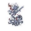
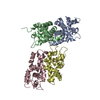
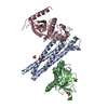
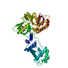
- PDB-3ty9: Crystal Structure of C. Thermocellum PNKP Ligase Domain AMP-Adenylate -
+ Open data

- Basic information
| Entry | Database: PDB / ID: 3ty9 | ||||||
|---|---|---|---|---|---|---|---|
| Title | Crystal Structure of C. Thermocellum PNKP Ligase Domain AMP-Adenylate | ||||||
 Components Components | Polynucleotide 2',3'-cyclic phosphate phosphodiesterase / polynucleotide 5'-hydroxyl-kinase / polynucleotide 3'-phosphatase | ||||||
 Keywords Keywords | TRANSFERASE / DNA ligase/mRNA capping enzyme / RNA Ligase / Adenylyltransferase / HEN1 | ||||||
| Function / homology |  Function and homology information Function and homology informationphosphatase activity / GTP binding / ATP binding / metal ion binding / cytoplasm Similarity search - Function | ||||||
| Biological species |  Clostridium thermocellum (bacteria) Clostridium thermocellum (bacteria) | ||||||
| Method |  X-RAY DIFFRACTION / SYNCHROTRON / MOLECULAR REPLACEMENT / Resolution: 3.12 Å X-RAY DIFFRACTION / SYNCHROTRON / MOLECULAR REPLACEMENT / Resolution: 3.12 Å | ||||||
 Authors Authors | Smith, P. / Wang, L. / Shuman, S. | ||||||
 Citation Citation | Journal: Proc.Natl.Acad.Sci.USA / Year: 2012 Title: The adenylyltransferase domain of bacterial Pnkp defines a unique RNA ligase family. Authors: Smith, P. / Wang, L.K. / Nair, P.A. / Shuman, S. | ||||||
| History |
|
- Structure visualization
| Structure viewer | Molecule: MolmilJmol/JSmol |
|---|
- Downloads & links
-Download
| PDBx/mmCIF format | 3ty9.cif.gz | 312.7 KB | Display | PDBx/mmCIF format |
|---|---|---|---|---|
| PDB format | pdb3ty9.ent.gz | 252.6 KB | Display | PDB format |
| PDBx/mmJSON format | 3ty9.json.gz | Tree view | PDBx/mmJSON format | |
| Others |  Other downloads Other downloads |
-Validation report
| Arichive directory | https://data.pdbj.org/pub/pdb/validation_reports/ty/3ty9ftp://data.pdbj.org/pub/pdb/validation_reports/ty/3ty9 | HTTPS FTP |
|---|
-Related structure data
| Related structure data |  3ty5SC  3ty8C S: Starting model for refinement C: citing same article ( |
|---|---|
| Similar structure data |


-Links
 PDBj
PDBj


- Assembly
| Deposited unit | 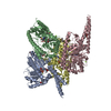
| ||||||||
|---|---|---|---|---|---|---|---|---|---|
| 1 | 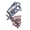
| ||||||||
| 2 | 
| ||||||||
| 3 | 
| ||||||||
| 4 | 
| ||||||||
| Unit cell |
| ||||||||
| Details | PROTEIN IS KNOWN TO FORM A DIMER WHICH INTERACTS WITH A DIMER OF HEN1 TO GIVE A HETEREOTETRAMER (2-PNKP/2-HEN1). THE OLIGOMERIC ORGANIZATIONS PRESENT IN THE CRYSTAL ARE ENUMERATED BELOW BUT MAY NOT CORRESPOND TO ANY OLIGOMER PRESENT IN THE HEN1/PNKP HETERODIMER |
-Components
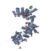
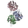
-Protein , 1 types, 4 molecules ABCD
| #1: Protein | Mass: 45100.680 Da / Num. of mol.: 4 / Fragment: Nucleotide Ligase Source method: isolated from a genetically manipulated source Source: (gene. exp.) Clostridium thermocellum (bacteria) / Strain: ATCC 27405 / Gene: Cthe_2768 / Plasmid: pET28b-smt3 / Production host: |
|---|
-Non-polymers , 6 types, 77 molecules 


| #2: Chemical | ChemComp-AMP /  Mass: 347.221 Da / Num. of mol.: 4 / Source method: obtained synthetically / Formula: C10H14N5O7P / Comment: AMP*YM Mass: 347.221 Da / Num. of mol.: 4 / Source method: obtained synthetically / Formula: C10H14N5O7P / Comment: AMP*YM#3: Chemical | ChemComp-MPD / (  Mass: 118.174 Da / Num. of mol.: 10 / Source method: obtained synthetically / Formula: C6H14O2 / Comment: precipitant*YM Mass: 118.174 Da / Num. of mol.: 10 / Source method: obtained synthetically / Formula: C6H14O2 / Comment: precipitant*YM#4: Chemical | ChemComp-MRD / (  Mass: 118.174 Da / Num. of mol.: 6 / Source method: obtained synthetically / Formula: C6H14O2 / Comment: precipitant*YM Mass: 118.174 Da / Num. of mol.: 6 / Source method: obtained synthetically / Formula: C6H14O2 / Comment: precipitant*YM#5: Chemical | ChemComp-GOL / |  Mass: 92.094 Da / Num. of mol.: 1 / Source method: obtained synthetically / Formula: C3H8O3 Mass: 92.094 Da / Num. of mol.: 1 / Source method: obtained synthetically / Formula: C3H8O3#6: Chemical | ChemComp-MG /  Mass: 24.305 Da / Num. of mol.: 4 / Source method: obtained synthetically / Formula: Mg Mass: 24.305 Da / Num. of mol.: 4 / Source method: obtained synthetically / Formula: Mg#7: Water | ChemComp-HOH / | Mass: 18.015 Da / Num. of mol.: 52 / Source method: isolated from a natural source / Formula: H2O |
|---|
-Details
| Has protein modification | Y |
|---|
-Experimental details
-Experiment
| Experiment | Method: X-RAY DIFFRACTION / Number of used crystals: 1 |
|---|
- Sample preparation
| Crystal | Density Matthews: 2.86 Å3/Da / Density % sol: 56.99 % |
|---|---|
| Crystal grow | Temperature: 295 K / Method: vapor diffusion, sitting drop / pH: 7.1 Details: HEPES buffer 20% (v/v) Hexylene Glycol, pH 7.1, VAPOR DIFFUSION, SITTING DROP, temperature 295K |
-Data collection
| Diffraction | Mean temperature: 130 K |
|---|---|
| Diffraction source | Source: SYNCHROTRON / Site: NSLS  / Beamline: X25 / Wavelength: 1.1 Å / Beamline: X25 / Wavelength: 1.1 Å |
| Detector | Type: DECTRIS PILATUS 6M / Detector: PIXEL / Date: Feb 15, 2011 / Details: See Beamline Documentation |
| Radiation | Monochromator: See Beamline Documentation / Protocol: SINGLE WAVELENGTH / Monochromatic (M) / Laue (L): M / Scattering type: x-ray |
| Radiation wavelength | Wavelength: 1.1 Å / Relative weight: 1 |
| Reflection | Resolution: 3.12→50 Å / Num. all: 37602 / Num. obs: 37527 / % possible obs: 99.8 % / Observed criterion σ(I): -3 / Redundancy: 7.3 % / Rsym value: 0.065 / Net I/σ(I): 9.2 |
- Processing
| Software |
| |||||||||||||||||||||||||||||||||||||||||||||||||||||||||||||||||||||||||||||||||||||||||||
|---|---|---|---|---|---|---|---|---|---|---|---|---|---|---|---|---|---|---|---|---|---|---|---|---|---|---|---|---|---|---|---|---|---|---|---|---|---|---|---|---|---|---|---|---|---|---|---|---|---|---|---|---|---|---|---|---|---|---|---|---|---|---|---|---|---|---|---|---|---|---|---|---|---|---|---|---|---|---|---|---|---|---|---|---|---|---|---|---|---|---|---|---|
| Refinement | Method to determine structure: MOLECULAR REPLACEMENT Starting model: PDB 3TY5 Resolution: 3.12→41.185 Å / SU ML: 0.38 / σ(F): 1.38 / Phase error: 31.99 / Stereochemistry target values: ML
| |||||||||||||||||||||||||||||||||||||||||||||||||||||||||||||||||||||||||||||||||||||||||||
| Solvent computation | Shrinkage radii: 0.83 Å / VDW probe radii: 1.1 Å / Solvent model: FLAT BULK SOLVENT MODEL / Bsol: 35.301 Å2 / ksol: 0.263 e/Å3 | |||||||||||||||||||||||||||||||||||||||||||||||||||||||||||||||||||||||||||||||||||||||||||
| Displacement parameters |
| |||||||||||||||||||||||||||||||||||||||||||||||||||||||||||||||||||||||||||||||||||||||||||
| Refinement step | Cycle: LAST / Resolution: 3.12→41.185 Å
| |||||||||||||||||||||||||||||||||||||||||||||||||||||||||||||||||||||||||||||||||||||||||||
| Refine LS restraints |
| |||||||||||||||||||||||||||||||||||||||||||||||||||||||||||||||||||||||||||||||||||||||||||
| LS refinement shell |
|
