 Movie
Movie Controller

[English] 日本語
 Yorodumi
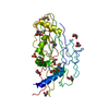
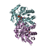
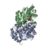
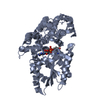
- PDB-1jfz: Crystal Structure of MN(II)-Complex of RNAse III Endonuclease Dom... -
+ Open data

- Basic information
| Entry | Database: PDB / ID: 1jfz | ||||||
|---|---|---|---|---|---|---|---|
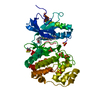
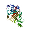
| Title | Crystal Structure of MN(II)-Complex of RNAse III Endonuclease Domain from Aquifex Aeolicus at 2.10 Angstrom Resolution | ||||||
 Components Components | RIBONUCLEASE III | ||||||
 Keywords Keywords | HYDROLASE / RIBONUCLEASE / RNASE III / DOUBLE-STRANDED RNA / RNA INTERFERENCE / ENDONUCLEASE DOMAIN / ENDONUCLEOLYTIC CLEAVAGE | ||||||
| Function / homology |  Function and homology information Function and homology informationribonuclease III / ribonuclease III activity / tRNA processing / RNA processing / mRNA processing / rRNA processing / double-stranded RNA binding / regulation of gene expression / metal ion binding / identical protein binding / cytosol Similarity search - Function | ||||||
| Biological species |   Aquifex aeolicus (bacteria) Aquifex aeolicus (bacteria) | ||||||
| Method |  X-RAY DIFFRACTION / SYNCHROTRON / MOLECULAR REPLACEMENT / Resolution: 2.1 Å X-RAY DIFFRACTION / SYNCHROTRON / MOLECULAR REPLACEMENT / Resolution: 2.1 Å | ||||||
 Authors Authors | Blaszczyk, J. / Ji, X. | ||||||
 Citation Citation | Journal: Structure / Year: 2001 Title: Crystallographic and modeling studies of RNase III suggest a mechanism for double-stranded RNA cleavage. Authors: Blaszczyk, J. / Tropea, J.E. / Bubunenko, M. / Routzahn, K.M. / Waugh, D.S. / Court, D.L. / Ji, X. | ||||||
| History |
|
- Structure visualization
| Structure viewer | Molecule: MolmilJmol/JSmol |
|---|
- Downloads & links
-Download
| PDBx/mmCIF format | 1jfz.cif.gz | 153.5 KB | Display | PDBx/mmCIF format |
|---|---|---|---|---|
| PDB format | pdb1jfz.ent.gz | 117.7 KB | Display | PDB format |
| PDBx/mmJSON format | 1jfz.json.gz | Tree view | PDBx/mmJSON format | |
| Others |  Other downloads Other downloads |
-Validation report
| Arichive directory | https://data.pdbj.org/pub/pdb/validation_reports/jf/1jfzftp://data.pdbj.org/pub/pdb/validation_reports/jf/1jfz | HTTPS FTP |
|---|
-Related structure data
| Related structure data | 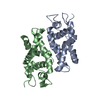 1i4sSC S: Starting model for refinement C: citing same article ( |
|---|---|
| Similar structure data |


-Links
 PDBj
PDBj

- Assembly
| Deposited unit | 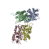
| ||||||||
|---|---|---|---|---|---|---|---|---|---|
| 1 | 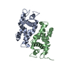
| ||||||||
| 2 | 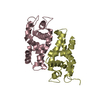
| ||||||||
| Unit cell |
| ||||||||
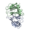
| Details | The biological assembly is a dimer. The asymmetric unit is composed from two independent dimers: first dimer is composed from identical chains A and B, and second dimer from identical chains C and D |
-Components
| #1: Protein | Mass: 18163.012 Da / Num. of mol.: 4 / Fragment: N-TERMINAL ENDONUCLEASE DOMAIN Source method: isolated from a genetically manipulated source Source: (gene. exp.) Aquifex aeolicus (bacteria) / Plasmid: PKM803 / Species (production host): Escherichia coli / Production host: #2: Chemical | ChemComp-MN /   Mass: 54.938 Da / Num. of mol.: 4 / Source method: obtained synthetically / Formula: Mn Mass: 54.938 Da / Num. of mol.: 4 / Source method: obtained synthetically / Formula: Mn#3: Water | ChemComp-HOH / |  Mass: 18.015 Da / Num. of mol.: 712 / Source method: isolated from a natural source / Formula: H2O Mass: 18.015 Da / Num. of mol.: 712 / Source method: isolated from a natural source / Formula: H2O |
|---|
-Experimental details
-Experiment
| Experiment | Method: X-RAY DIFFRACTION / Number of used crystals: 1 |
|---|
- Sample preparation
| Crystal | Density Matthews: 2.13 Å3/Da / Density % sol: 39.7 % | |||||||||||||||||||||||||||||||||||||||||||||||||
|---|---|---|---|---|---|---|---|---|---|---|---|---|---|---|---|---|---|---|---|---|---|---|---|---|---|---|---|---|---|---|---|---|---|---|---|---|---|---|---|---|---|---|---|---|---|---|---|---|---|---|
| Crystal grow | Temperature: 292 K / Method: vapor diffusion, hanging drop / pH: 8.5 Details: PEG4000, TRIS-HCL, ACETATE, CHLORIDE, pH 8.50, VAPOR DIFFUSION, HANGING DROP, temperature 292K | |||||||||||||||||||||||||||||||||||||||||||||||||
| Crystal grow | *PLUS Temperature: 18-20 ℃ / pH: 7.5 | |||||||||||||||||||||||||||||||||||||||||||||||||
| Components of the solutions | *PLUS
|
-Data collection
| Diffraction | Mean temperature: 100 K |
|---|---|
| Diffraction source | Source: SYNCHROTRON / Site: NSLS  / Beamline: X9B / Wavelength: 1.045 / Beamline: X9B / Wavelength: 1.045 |
| Detector | Type: ADSC QUANTUM 4 / Detector: CCD / Date: Mar 21, 2001 / Details: MIRROR |
| Radiation | Monochromator: SILICON 111 / Protocol: SINGLE WAVELENGTH / Monochromatic (M) / Laue (L): M / Scattering type: x-ray |
| Radiation wavelength | Wavelength: 1.045 Å / Relative weight: 1 |
| Reflection | Resolution: 2.1→30 Å / Num. all: 33591 / Num. obs: 33591 / % possible obs: 92.9 % / Observed criterion σ(F): 0 / Observed criterion σ(I): 0 / Redundancy: 2.165 % / Biso Wilson estimate: 28.3 Å2 / Rmerge(I) obs: 0.059 / Net I/σ(I): 10.3132 |
| Reflection shell | Resolution: 2.1→2.18 Å / Redundancy: 2.023 % / Rmerge(I) obs: 0.185 / Mean I/σ(I) obs: 2.985 / Num. unique all: 2721 / % possible all: 74.7 |
| Reflection | *PLUS Num. measured all: 72740 |
| Reflection shell | *PLUS % possible obs: 74.7 % / Mean I/σ(I) obs: 3 |
- Processing
| Software |
| ||||||||||||||||||||||||||||||||||||||||||||
|---|---|---|---|---|---|---|---|---|---|---|---|---|---|---|---|---|---|---|---|---|---|---|---|---|---|---|---|---|---|---|---|---|---|---|---|---|---|---|---|---|---|---|---|---|---|
| Refinement | Method to determine structure: MOLECULAR REPLACEMENT Starting model: PDB ID 1I4S Resolution: 2.1→30 Å / Num. parameters: 21898 / Num. restraintsaints: 20454 / Isotropic thermal model: ISOTROPIC / Cross valid method: FREE R / σ(F): 4 / σ(I): 2 / Stereochemistry target values: ENGH AND HUBER Details: LEAST-SQUARES REFINEMENT USING THE KONNERT-HENDRICKSON CONJUGATE-GRADIENT ALGORITHM
| ||||||||||||||||||||||||||||||||||||||||||||
| Solvent computation | Solvent model: MOEWS & KRETSINGER, J.MOL.BIOL.91(1975) 201-228 | ||||||||||||||||||||||||||||||||||||||||||||
| Displacement parameters | Biso mean: 31.865 Å2 | ||||||||||||||||||||||||||||||||||||||||||||
| Refine analyze | Luzzati coordinate error obs: 0.228 Å / Luzzati d res low obs: 5 Å / Num. disordered residues: 7 / Occupancy sum hydrogen: 0 / Occupancy sum non hydrogen: 5640 | ||||||||||||||||||||||||||||||||||||||||||||
| Refinement step | Cycle: LAST / Resolution: 2.1→30 Å
| ||||||||||||||||||||||||||||||||||||||||||||
| Refine LS restraints |
| ||||||||||||||||||||||||||||||||||||||||||||
| LS refinement shell |
| ||||||||||||||||||||||||||||||||||||||||||||
| Software | *PLUS Name: SHELXL-97 / Classification: refinement | ||||||||||||||||||||||||||||||||||||||||||||
| Refinement | *PLUS σ(F): 4 / Rfactor all: 0.209 / Rfactor obs: 0.191 / Rfactor Rfree: 0.277 | ||||||||||||||||||||||||||||||||||||||||||||
| Solvent computation | *PLUS | ||||||||||||||||||||||||||||||||||||||||||||
| Displacement parameters | *PLUS | ||||||||||||||||||||||||||||||||||||||||||||
| Refine LS restraints | *PLUS
|
