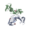
Entry Database : PDB / ID : 3tx7Title Crystal structure of LRH-1/beta-catenin complex Catenin beta-1 Nuclear receptor subfamily 5 group A member 2 Keywords / / / / Function / homology Function Domain/homology Component
/ / / / / / / / / / / / / / / / / / / / / / / / / / / / / / / / / / / / / / / / / / / / / / / / / / / / / / / / / / / / / / / / / / / / / / / / / / / / / / / / / / / / / / / / / / / / / / / / / / / / / / / / / / / / / / / / / / / / / / / / / / / / / / / / / / / / / / / / / / / / / / / Biological species Homo sapiens (human)Method / / / Resolution : 2.76 Å Authors Yumoto, F. / Fletterick, R. Journal : Proc.Natl.Acad.Sci.USA / Year : 2012Title : Structural basis of coactivation of liver receptor homolog-1 by beta-catenin.Authors : Yumoto, F. / Nguyen, P. / Sablin, E.P. / Baxter, J.D. / Webb, P. / Fletterick, R.J. History Deposition Sep 22, 2011 Deposition site / Processing site Revision 1.0 Dec 14, 2011 Provider / Type Revision 1.1 Dec 28, 2011 Group Revision 1.2 Jun 19, 2013 Group Revision 1.3 Sep 13, 2023 Group Data collection / Database references ... Data collection / Database references / Derived calculations / Refinement description Category chem_comp_atom / chem_comp_bond ... chem_comp_atom / chem_comp_bond / database_2 / pdbx_initial_refinement_model / struct_ref_seq_dif / struct_site Item _database_2.pdbx_DOI / _database_2.pdbx_database_accession ... _database_2.pdbx_DOI / _database_2.pdbx_database_accession / _struct_ref_seq_dif.details / _struct_site.pdbx_auth_asym_id / _struct_site.pdbx_auth_comp_id / _struct_site.pdbx_auth_seq_id
Show all Show less
 Movie
Movie Controller


 Open data
 Basic information
 Components
 Keywords
 Function and homology information
 Homo sapiens (human)
 X-RAY DIFFRACTION /
  Authors
 Citation
 Structure visualization
 Downloads & links
 Other downloads


 PDBj
PDBj


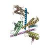
 Assembly
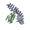

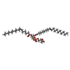
 Mass: 746.991 Da / Num. of mol.: 1 / Source method: obtained synthetically / Formula: C40H75O10P / Comment: phospholipid*YM
 Sample preparation
 / Beamline: 8.3.1 / Wavelength: 1.11587 Å
 Processing