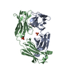
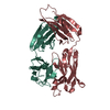
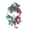
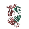
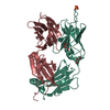
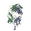
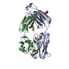
登録情報 データベース : PDB / ID : 3tv3タイトル Crystal structure of broad and potent HIV-1 neutralizing antibody PGT128 in complex with Man9 PGT128 heavy chain, Ig gamma-1 chain C region PGT128 light chain, Ig lambda-2 chain C regions キーワード / / / 機能・相同性 分子機能 ドメイン・相同性 構成要素
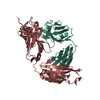
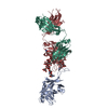
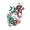
/ / / / / / / / / / / / / / / / / / / / / / / / / / / / / / / / / / / / / / / / / / / / / / / / / / / / / / / / / / / / / / / / / / / / / / / / / / / / / 生物種 Homo sapiens (ヒト)手法 / / / 解像度 : 1.29 Å データ登録者 Pejchal, R. / Wilson, I.A. ジャーナル : Science / 年 : 2011タイトル : A potent and broad neutralizing antibody recognizes and penetrates the HIV glycan shield.著者: Robert Pejchal / Katie J Doores / Laura M Walker / Reza Khayat / Po-Ssu Huang / Sheng-Kai Wang / Robyn L Stanfield / Jean-Philippe Julien / Alejandra Ramos / Max Crispin / Rafael Depetris / ... 著者 : Robert Pejchal / Katie J Doores / Laura M Walker / Reza Khayat / Po-Ssu Huang / Sheng-Kai Wang / Robyn L Stanfield / Jean-Philippe Julien / Alejandra Ramos / Max Crispin / Rafael Depetris / Umesh Katpally / Andre Marozsan / Albert Cupo / Sebastien Maloveste / Yan Liu / Ryan McBride / Yukishige Ito / Rogier W Sanders / Cassandra Ogohara / James C Paulson / Ten Feizi / Christopher N Scanlan / Chi-Huey Wong / John P Moore / William C Olson / Andrew B Ward / Pascal Poignard / William R Schief / Dennis R Burton / Ian A Wilson / 要旨 : The HIV envelope (Env) protein gp120 is protected from antibody recognition by a dense glycan shield. However, several of the recently identified PGT broadly neutralizing antibodies appear to ... The HIV envelope (Env) protein gp120 is protected from antibody recognition by a dense glycan shield. However, several of the recently identified PGT broadly neutralizing antibodies appear to interact directly with the HIV glycan coat. Crystal structures of antigen-binding fragments (Fabs) PGT 127 and 128 with Man(9) at 1.65 and 1.29 angstrom resolution, respectively, and glycan binding data delineate a specific high mannose-binding site. Fab PGT 128 complexed with a fully glycosylated gp120 outer domain at 3.25 angstroms reveals that the antibody penetrates the glycan shield and recognizes two conserved glycans as well as a short β-strand segment of the gp120 V3 loop, accounting for its high binding affinity and broad specificity. Furthermore, our data suggest that the high neutralization potency of PGT 127 and 128 immunoglobulin Gs may be mediated by cross-linking Env trimers on the viral surface. 履歴 登録 2011年9月19日 登録サイト / 処理サイト 改定 1.0 2011年10月19日 Provider / タイプ 改定 1.1 2011年10月26日 Group 改定 1.2 2011年12月7日 Group 改定 1.3 2017年8月2日 Group / Source and taxonomy / カテゴリ / software改定 2.0 2019年12月25日 Group Advisory / Database references ... Advisory / Database references / Derived calculations / Polymer sequence カテゴリ entity_poly / pdbx_struct_mod_residue ... entity_poly / pdbx_struct_mod_residue / pdbx_unobs_or_zero_occ_atoms / struct_conn / struct_ref_seq Item _entity_poly.pdbx_seq_one_letter_code_can / _pdbx_struct_mod_residue.parent_comp_id ... _entity_poly.pdbx_seq_one_letter_code_can / _pdbx_struct_mod_residue.parent_comp_id / _struct_conn.pdbx_leaving_atom_flag / _struct_ref_seq.db_align_end 改定 3.0 2020年7月29日 Group Atomic model / Data collection ... Atomic model / Data collection / Derived calculations / Structure summary カテゴリ atom_site / atom_site_anisotrop ... atom_site / atom_site_anisotrop / chem_comp / entity / pdbx_branch_scheme / pdbx_chem_comp_identifier / pdbx_entity_branch / pdbx_entity_branch_descriptor / pdbx_entity_branch_link / pdbx_entity_branch_list / pdbx_entity_nonpoly / pdbx_nonpoly_scheme / pdbx_struct_assembly_gen / struct_asym / struct_conn / struct_site / struct_site_gen Item _atom_site.B_iso_or_equiv / _atom_site.Cartn_x ... _atom_site.B_iso_or_equiv / _atom_site.Cartn_x / _atom_site.Cartn_y / _atom_site.Cartn_z / _atom_site.auth_asym_id / _atom_site.auth_seq_id / _atom_site.label_asym_id / _atom_site_anisotrop.U[1][1] / _atom_site_anisotrop.U[1][2] / _atom_site_anisotrop.U[1][3] / _atom_site_anisotrop.U[2][2] / _atom_site_anisotrop.U[2][3] / _atom_site_anisotrop.U[3][3] / _atom_site_anisotrop.pdbx_auth_asym_id / _atom_site_anisotrop.pdbx_auth_seq_id / _atom_site_anisotrop.pdbx_label_asym_id / _chem_comp.name / _chem_comp.type / _entity.formula_weight / _entity.pdbx_description / _entity.pdbx_number_of_molecules / _entity.type / _pdbx_struct_assembly_gen.asym_id_list / _struct_conn.pdbx_dist_value / _struct_conn.ptnr1_auth_asym_id / _struct_conn.ptnr1_auth_comp_id / _struct_conn.ptnr1_auth_seq_id / _struct_conn.ptnr1_label_asym_id / _struct_conn.ptnr1_label_atom_id / _struct_conn.ptnr1_label_comp_id / _struct_conn.ptnr2_auth_asym_id / _struct_conn.ptnr2_auth_comp_id / _struct_conn.ptnr2_auth_seq_id / _struct_conn.ptnr2_label_asym_id / _struct_conn.ptnr2_label_atom_id / _struct_conn.ptnr2_label_comp_id 解説 / Provider / タイプ 改定 3.1 2021年4月28日 Group / Source and taxonomy / Structure summaryカテゴリ / entity_src_gen / struct_connItem _chem_comp.pdbx_synonyms / _entity_src_gen.host_org_common_name ... _chem_comp.pdbx_synonyms / _entity_src_gen.host_org_common_name / _entity_src_gen.pdbx_host_org_cell_line / _entity_src_gen.pdbx_host_org_strain / _struct_conn.pdbx_leaving_atom_flag 改定 3.2 2024年10月30日 Group / Database references / Structure summaryカテゴリ chem_comp_atom / chem_comp_bond ... chem_comp_atom / chem_comp_bond / database_2 / pdbx_entry_details / pdbx_modification_feature Item / _database_2.pdbx_database_accession
すべて表示 表示を減らす
 ムービー
ムービー コントローラー


 データを開く
 基本情報
基本情報 要素
要素 キーワード
キーワード 機能・相同性情報
 Homo sapiens (ヒト)
 X線回折 /
  データ登録者
 引用
引用
 構造の表示
構造の表示 ダウンロードとリンク
 その他のダウンロード
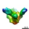


 PDBj
PDBj


 集合体
集合体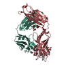


 分子量: 92.094 Da / 分子数: 4 / 由来タイプ: 合成 / 式: C3H8O3
 分子量: 87.163 Da / 分子数: 1 / 由来タイプ: 合成 / 式: C5H13N
 分子量: 238.305 Da / 分子数: 1 / 由来タイプ: 合成 / 式: C8H18N2O4S / コメント: pH緩衝剤*YM
 試料調製
試料調製 解析
解析