 Movie
Movie Controller

[English] 日本語
 Yorodumi
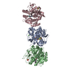
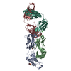
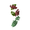
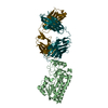
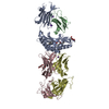
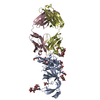
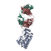
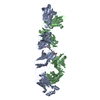
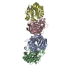
- PDB-3tyg: Crystal structure of broad and potent HIV-1 neutralizing antibody... -
+ Open data

- Basic information
| Entry | Database: PDB / ID: 3tyg | ||||||||||||
|---|---|---|---|---|---|---|---|---|---|---|---|---|---|
| Title | Crystal structure of broad and potent HIV-1 neutralizing antibody PGT128 in complex with a glycosylated engineered gp120 outer domain with miniV3 (eODmV3) | ||||||||||||
 Components Components |
| ||||||||||||
 Keywords Keywords | IMMUNE SYSTEM / VIRAL PROTEIN / gp120 / HIV-1 / Env / Fab / HIV-1 neutralizing antibody | ||||||||||||
| Function / homology |  Function and homology information Function and homology informationIgD immunoglobulin complex / positive regulation of B cell activation / IgA immunoglobulin complex / IgM immunoglobulin complex / phagocytosis, recognition / IgE immunoglobulin complex / Synthesis and processing of ENV and VPU / symbiont-mediated evasion of host immune response / CD22 mediated BCR regulation / positive regulation of establishment of T cell polarity ...IgD immunoglobulin complex / positive regulation of B cell activation / IgA immunoglobulin complex / IgM immunoglobulin complex / phagocytosis, recognition / IgE immunoglobulin complex / Synthesis and processing of ENV and VPU / symbiont-mediated evasion of host immune response / CD22 mediated BCR regulation / positive regulation of establishment of T cell polarity / Alpha-defensins / complement-dependent cytotoxicity / antibody-dependent cellular cytotoxicity / Fc-gamma receptor I complex binding / Fc epsilon receptor (FCERI) signaling / immunoglobulin complex, circulating / Classical antibody-mediated complement activation / phagocytosis, engulfment / immunoglobulin receptor binding / Initial triggering of complement / IgG immunoglobulin complex / Dectin-2 family / immunoglobulin mediated immune response / FCGR activation / Role of LAT2/NTAL/LAB on calcium mobilization / complement activation, classical pathway / Binding and entry of HIV virion / Role of phospholipids in phagocytosis / Scavenging of heme from plasma / antigen binding / symbiont-mediated perturbation of host defense response / positive regulation of plasma membrane raft polarization / positive regulation of receptor clustering / FCERI mediated Ca+2 mobilization / FCGR3A-mediated IL10 synthesis / Regulation of Complement cascade / Antigen activates B Cell Receptor (BCR) leading to generation of second messengers / host cell endosome membrane / Cell surface interactions at the vascular wall / actin filament organization / B cell receptor signaling pathway / FCGR3A-mediated phagocytosis / FCERI mediated MAPK activation / Assembly Of The HIV Virion / Budding and maturation of HIV virion / Regulation of actin dynamics for phagocytic cup formation / FCERI mediated NF-kB activation / Immunoregulatory interactions between a Lymphoid and a non-Lymphoid cell / antibacterial humoral response / Interleukin-4 and Interleukin-13 signaling / blood microparticle / clathrin-dependent endocytosis of virus by host cell / Potential therapeutics for SARS / adaptive immune response / defense response to bacterium / viral protein processing / receptor ligand activity / fusion of virus membrane with host plasma membrane / external side of plasma membrane / innate immune response / fusion of virus membrane with host endosome membrane / viral envelope / symbiont entry into host cell / virion attachment to host cell / host cell plasma membrane / virion membrane / structural molecule activity / : / extracellular exosome / extracellular region / membrane / identical protein binding / plasma membrane Similarity search - Function | ||||||||||||
| Biological species |   Human immunodeficiency virus 1 Human immunodeficiency virus 1 Homo sapiens (human) Homo sapiens (human) | ||||||||||||
| Method |  X-RAY DIFFRACTION / SYNCHROTRON / MOLECULAR REPLACEMENT / Resolution: 3.25 Å X-RAY DIFFRACTION / SYNCHROTRON / MOLECULAR REPLACEMENT / Resolution: 3.25 Å | ||||||||||||
 Authors Authors | Pejchal, R. / Huang, P.S. / Schief, W.R. / Stanfield, R.L. / Wilson, I.A. | ||||||||||||
 Citation Citation | Journal: Science / Year: 2011 Title: A potent and broad neutralizing antibody recognizes and penetrates the HIV glycan shield. Authors: Robert Pejchal / Katie J Doores / Laura M Walker / Reza Khayat / Po-Ssu Huang / Sheng-Kai Wang / Robyn L Stanfield / Jean-Philippe Julien / Alejandra Ramos / Max Crispin / Rafael Depetris / ...Authors: Robert Pejchal / Katie J Doores / Laura M Walker / Reza Khayat / Po-Ssu Huang / Sheng-Kai Wang / Robyn L Stanfield / Jean-Philippe Julien / Alejandra Ramos / Max Crispin / Rafael Depetris / Umesh Katpally / Andre Marozsan / Albert Cupo / Sebastien Maloveste / Yan Liu / Ryan McBride / Yukishige Ito / Rogier W Sanders / Cassandra Ogohara / James C Paulson / Ten Feizi / Christopher N Scanlan / Chi-Huey Wong / John P Moore / William C Olson / Andrew B Ward / Pascal Poignard / William R Schief / Dennis R Burton / Ian A Wilson /  Abstract: The HIV envelope (Env) protein gp120 is protected from antibody recognition by a dense glycan shield. However, several of the recently identified PGT broadly neutralizing antibodies appear to ...The HIV envelope (Env) protein gp120 is protected from antibody recognition by a dense glycan shield. However, several of the recently identified PGT broadly neutralizing antibodies appear to interact directly with the HIV glycan coat. Crystal structures of antigen-binding fragments (Fabs) PGT 127 and 128 with Man(9) at 1.65 and 1.29 angstrom resolution, respectively, and glycan binding data delineate a specific high mannose-binding site. Fab PGT 128 complexed with a fully glycosylated gp120 outer domain at 3.25 angstroms reveals that the antibody penetrates the glycan shield and recognizes two conserved glycans as well as a short β-strand segment of the gp120 V3 loop, accounting for its high binding affinity and broad specificity. Furthermore, our data suggest that the high neutralization potency of PGT 127 and 128 immunoglobulin Gs may be mediated by cross-linking Env trimers on the viral surface. | ||||||||||||
| History |
|
- Structure visualization
| Structure viewer | Molecule: MolmilJmol/JSmol |
|---|
- Downloads & links
-Download
| PDBx/mmCIF format | 3tyg.cif.gz | 252.6 KB | Display | PDBx/mmCIF format |
|---|---|---|---|---|
| PDB format | pdb3tyg.ent.gz | 203.2 KB | Display | PDB format |
| PDBx/mmJSON format | 3tyg.json.gz | Tree view | PDBx/mmJSON format | |
| Others |  Other downloads Other downloads |
-Validation report
| Arichive directory | https://data.pdbj.org/pub/pdb/validation_reports/ty/3tygftp://data.pdbj.org/pub/pdb/validation_reports/ty/3tyg | HTTPS FTP |
|---|
-Related structure data
| Related structure data | 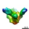 1970C 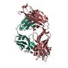 3tv3SC 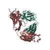 3twcC S: Starting model for refinement C: citing same article ( |
|---|---|
| Similar structure data |

-Links
 PDBj
PDBj


- Assembly
| Deposited unit | 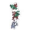
| ||||||||
|---|---|---|---|---|---|---|---|---|---|
| 1 |
| ||||||||
| Unit cell |
|
-Components
| #1: Protein | Mass: 22019.486 Da / Num. of mol.: 1 Source method: isolated from a genetically manipulated source Source: (gene. exp.) Human immunodeficiency virus 1 / Strain: HXB2, JR-FL / Gene: env / Plasmid: pLSEC / Cell line (production host): HEK 293S GnTI -/- / Production host: Homo sapiens (human) / References: UniProt: P04578, UniProt: Q75760 |
|---|---|
| #2: Antibody | Mass: 22206.559 Da / Num. of mol.: 1 Source method: isolated from a genetically manipulated source Source: (gene. exp.) Homo sapiens (human) / Gene: IGLC2 / Plasmid: pTT5 / Cell line (production host): HEK 293F / Production host: Homo sapiens (human) / References: UniProt: P0CG05, UniProt: P0DOY3*PLUS |
| #3: Antibody | Mass: 25562.686 Da / Num. of mol.: 1 Source method: isolated from a genetically manipulated source Source: (gene. exp.) Homo sapiens (human) / Gene: IGHG1 / Plasmid: pTT5 / Cell line (production host): HEK 293F / Production host: Homo sapiens (human) / References: UniProt: P01857 |
| #4: Polysaccharide | alpha-D-mannopyranose-(1-2)-alpha-D-mannopyranose-(1-2)-alpha-D-mannopyranose-(1-3)-[alpha-D- ...alpha-D-mannopyranose-(1-2)-alpha-D-mannopyranose-(1-2)-alpha-D-mannopyranose-(1-3)-[alpha-D-mannopyranose-(1-2)-alpha-D-mannopyranose-(1-6)-[alpha-D-mannopyranose-(1-3)]alpha-D-mannopyranose-(1-6)]beta-D-mannopyranose-(1-4)-2-acetamido-2-deoxy-beta-D-glucopyranose-(1-4)-2-acetamido-2-deoxy-beta-D-glucopyranose Source method: isolated from a genetically manipulated source |
| #5: Polysaccharide | alpha-D-mannopyranose-(1-3)-[alpha-D-mannopyranose-(1-6)]alpha-D-mannopyranose-(1-6)-[alpha-D- ...alpha-D-mannopyranose-(1-3)-[alpha-D-mannopyranose-(1-6)]alpha-D-mannopyranose-(1-6)-[alpha-D-mannopyranose-(1-3)]beta-D-mannopyranose-(1-4)-2-acetamido-2-deoxy-beta-D-glucopyranose-(1-4)-2-acetamido-2-deoxy-beta-D-glucopyranose Source method: isolated from a genetically manipulated source |
| Has protein modification | Y |
-Experimental details
-Experiment
| Experiment | Method: X-RAY DIFFRACTION / Number of used crystals: 1 |
|---|
- Sample preparation
| Crystal | Density Matthews: 3.23 Å3/Da / Density % sol: 61.91 % |
|---|---|
| Crystal grow | Temperature: 277 K / Method: vapor diffusion, sitting drop / pH: 8 Details: 24% PEG 3350, 0.29M CaCl2, pH 8.0, VAPOR DIFFUSION, SITTING DROP, temperature 277K |
-Data collection
| Diffraction | Mean temperature: 100 K |
|---|---|
| Diffraction source | Source: SYNCHROTRON / Site: SSRL / Beamline: BL11-1 / Wavelength: 0.97945 Å |
| Detector | Type: MARMOSAIC 325 mm CCD / Detector: CCD / Date: Jun 25, 2011 |
| Radiation | Monochromator: Side scattering bent cube-root I-beam single crystal Protocol: SINGLE WAVELENGTH / Monochromatic (M) / Laue (L): M / Scattering type: x-ray |
| Radiation wavelength | Wavelength: 0.97945 Å / Relative weight: 1 |
| Reflection | Resolution: 3.25→50 Å / Num. all: 14575 / Num. obs: 13988 / % possible obs: 91.7 % / Observed criterion σ(F): 0 / Observed criterion σ(I): 0 / Redundancy: 4.1 % / Biso Wilson estimate: 50.03 Å2 / Rsym value: 0.157 / Net I/σ(I): 6.6 |
| Reflection shell | Resolution: 3.25→3.37 Å / Mean I/σ(I) obs: 1.7 / Rsym value: 0.548 / % possible all: 55.6 |
- Processing
| Software |
| ||||||||||||||||||||||||||||||||||||||||||||||||||||||||||||||||||||||||||||||||||||||||||||||||||||||||||||||||||||||||||||||||||||||||||||||||||||||
|---|---|---|---|---|---|---|---|---|---|---|---|---|---|---|---|---|---|---|---|---|---|---|---|---|---|---|---|---|---|---|---|---|---|---|---|---|---|---|---|---|---|---|---|---|---|---|---|---|---|---|---|---|---|---|---|---|---|---|---|---|---|---|---|---|---|---|---|---|---|---|---|---|---|---|---|---|---|---|---|---|---|---|---|---|---|---|---|---|---|---|---|---|---|---|---|---|---|---|---|---|---|---|---|---|---|---|---|---|---|---|---|---|---|---|---|---|---|---|---|---|---|---|---|---|---|---|---|---|---|---|---|---|---|---|---|---|---|---|---|---|---|---|---|---|---|---|---|---|---|---|---|
| Refinement | Method to determine structure: MOLECULAR REPLACEMENT Starting model: PDB ENTRY 3TV3 Resolution: 3.25→49.7 Å / Cor.coef. Fo:Fc: 0.8398 / Cor.coef. Fo:Fc free: 0.7738 / Cross valid method: THROUGHOUT / σ(F): 0 / Stereochemistry target values: Engh & Huber
| ||||||||||||||||||||||||||||||||||||||||||||||||||||||||||||||||||||||||||||||||||||||||||||||||||||||||||||||||||||||||||||||||||||||||||||||||||||||
| Displacement parameters | Biso mean: 117.59 Å2
| ||||||||||||||||||||||||||||||||||||||||||||||||||||||||||||||||||||||||||||||||||||||||||||||||||||||||||||||||||||||||||||||||||||||||||||||||||||||
| Refine analyze | Luzzati coordinate error obs: 0.79 Å | ||||||||||||||||||||||||||||||||||||||||||||||||||||||||||||||||||||||||||||||||||||||||||||||||||||||||||||||||||||||||||||||||||||||||||||||||||||||
| Refinement step | Cycle: LAST / Resolution: 3.25→49.7 Å
| ||||||||||||||||||||||||||||||||||||||||||||||||||||||||||||||||||||||||||||||||||||||||||||||||||||||||||||||||||||||||||||||||||||||||||||||||||||||
| Refine LS restraints |
| ||||||||||||||||||||||||||||||||||||||||||||||||||||||||||||||||||||||||||||||||||||||||||||||||||||||||||||||||||||||||||||||||||||||||||||||||||||||
| LS refinement shell | Resolution: 3.25→3.51 Å / Total num. of bins used: 7
| ||||||||||||||||||||||||||||||||||||||||||||||||||||||||||||||||||||||||||||||||||||||||||||||||||||||||||||||||||||||||||||||||||||||||||||||||||||||
| Refinement TLS params. | Method: refined / Refine-ID: X-RAY DIFFRACTION
| ||||||||||||||||||||||||||||||||||||||||||||||||||||||||||||||||||||||||||||||||||||||||||||||||||||||||||||||||||||||||||||||||||||||||||||||||||||||
| Refinement TLS group |
|
