 Movie
Movie Controller

[English] 日本語
 Yorodumi
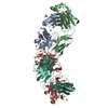
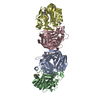
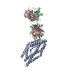
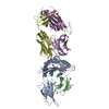
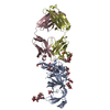
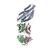
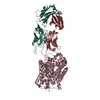
- PDB-4u6v: Mechanisms of Neutralization of a Human Anti-Alpha Toxin Antibody -
+ Open data

- Basic information
| Entry | Database: PDB / ID: 4u6v | |||||||||
|---|---|---|---|---|---|---|---|---|---|---|
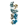
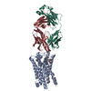
| Title | Mechanisms of Neutralization of a Human Anti-Alpha Toxin Antibody | |||||||||
 Components Components |
| |||||||||
 Keywords Keywords | IMMUNE SYSTEM / alpha toxin / Fab / Staphylococcus aureus / mutagenesis | |||||||||
| Function / homology |  Function and homology information Function and homology information | |||||||||
| Biological species |  Homo sapiens (human) Homo sapiens (human) Staphylococcus aureus subsp. aureus TCH60 (bacteria) Staphylococcus aureus subsp. aureus TCH60 (bacteria) | |||||||||
| Method |  X-RAY DIFFRACTION / SYNCHROTRON / MOLECULAR REPLACEMENT / Resolution: 2.56 Å X-RAY DIFFRACTION / SYNCHROTRON / MOLECULAR REPLACEMENT / Resolution: 2.56 Å | |||||||||
 Authors Authors | Oganesyan, V.Y. / Peng, L. / Damschroder, M.M. / Cheng, L. / Sadowska, A. / Tkaczyk, C. / Sellman, B. / Wu, H. / Dall'Acqua, W.F. | |||||||||
 Citation Citation | Journal: J.Biol.Chem. / Year: 2014 Title: Mechanisms of Neutralization of a Human Anti-alpha-toxin Antibody. Authors: Oganesyan, V. / Peng, L. / Damschroder, M.M. / Cheng, L. / Sadowska, A. / Tkaczyk, C. / Sellman, B.R. / Wu, H. / Dall'Acqua, W.F. | |||||||||
| History |
|
- Structure visualization
| Structure viewer | Molecule: MolmilJmol/JSmol |
|---|
- Downloads & links
-Download
| PDBx/mmCIF format | 4u6v.cif.gz | 561.5 KB | Display | PDBx/mmCIF format |
|---|---|---|---|---|
| PDB format | pdb4u6v.ent.gz | 465.8 KB | Display | PDB format |
| PDBx/mmJSON format | 4u6v.json.gz | Tree view | PDBx/mmJSON format | |
| Others |  Other downloads Other downloads |
-Validation report
| Arichive directory | https://data.pdbj.org/pub/pdb/validation_reports/u6/4u6vftp://data.pdbj.org/pub/pdb/validation_reports/u6/4u6v | HTTPS FTP |
|---|
-Related structure data
| Related structure data | 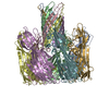 7ahlS S: Starting model for refinement |
|---|---|
| Similar structure data |

-Links
 PDBj
PDBj


- Assembly
| Deposited unit | 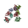
| |||||||||||||||||||||||||||||||||||||||||||||||||||||||||||||||||||||||||||||||||||||||||||||||
|---|---|---|---|---|---|---|---|---|---|---|---|---|---|---|---|---|---|---|---|---|---|---|---|---|---|---|---|---|---|---|---|---|---|---|---|---|---|---|---|---|---|---|---|---|---|---|---|---|---|---|---|---|---|---|---|---|---|---|---|---|---|---|---|---|---|---|---|---|---|---|---|---|---|---|---|---|---|---|---|---|---|---|---|---|---|---|---|---|---|---|---|---|---|---|---|---|
| 1 | 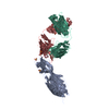
| |||||||||||||||||||||||||||||||||||||||||||||||||||||||||||||||||||||||||||||||||||||||||||||||
| 2 | 
| |||||||||||||||||||||||||||||||||||||||||||||||||||||||||||||||||||||||||||||||||||||||||||||||
| Unit cell |
| |||||||||||||||||||||||||||||||||||||||||||||||||||||||||||||||||||||||||||||||||||||||||||||||
| Noncrystallographic symmetry (NCS) | NCS domain:
NCS domain segments: Component-ID: _ / Refine code: _
NCS ensembles :
|
-Components
| #1: Antibody | Mass: 23794.520 Da / Num. of mol.: 2 Source method: isolated from a genetically manipulated source Source: (gene. exp.) Homo sapiens (human) / Production host: Mammalian expression vector pBGSA (others)#2: Antibody | Mass: 23404.002 Da / Num. of mol.: 2 Source method: isolated from a genetically manipulated source Source: (gene. exp.) Homo sapiens (human) / Production host: Mammalian expression vector pBGSA (others)#3: Protein | Mass: 33302.055 Da / Num. of mol.: 2 Source method: isolated from a genetically manipulated source Source: (gene. exp.) Staphylococcus aureus subsp. aureus TCH60 (bacteria)Production host:  Staphylococcus aureus (bacteria) / References: UniProt: Q2G1X0 Staphylococcus aureus (bacteria) / References: UniProt: Q2G1X0#4: Chemical | ChemComp-SO4 /   Mass: 96.063 Da / Num. of mol.: 6 / Source method: obtained synthetically / Formula: SO4 Mass: 96.063 Da / Num. of mol.: 6 / Source method: obtained synthetically / Formula: SO4#5: Water | ChemComp-HOH / |  Mass: 18.015 Da / Num. of mol.: 88 / Source method: isolated from a natural source / Formula: H2O Mass: 18.015 Da / Num. of mol.: 88 / Source method: isolated from a natural source / Formula: H2OHas protein modification | Y | |
|---|
-Experimental details
-Experiment
| Experiment | Method: X-RAY DIFFRACTION / Number of used crystals: 1 |
|---|
- Sample preparation
| Crystal | Density Matthews: 3.5 Å3/Da / Density % sol: 64.8 % |
|---|---|
| Crystal grow | Temperature: 293 K / Method: vapor diffusion, hanging drop / pH: 7.5 Details: Diffraction quality crystals grew in about a week in hanging drops where 3 ul of the MEDI4893 Fab/AT complex at a concentration of 15 mg/ml in 50 mM Tris-HCl, pH 7.5, 100 mM NaCl was mixed ...Details: Diffraction quality crystals grew in about a week in hanging drops where 3 ul of the MEDI4893 Fab/AT complex at a concentration of 15 mg/ml in 50 mM Tris-HCl, pH 7.5, 100 mM NaCl was mixed with 2 ul of 10% (w/v) PEG 6000, 100 mM HEPES, pH 7.0 and let to equilibrate against 300 ul of 10% (w/v) PEG 6000, 100 mM HEPES, pH 7.0 |
-Data collection
| Diffraction | Mean temperature: 100 K |
|---|---|
| Diffraction source | Source: SYNCHROTRON / Site: APS  / Beamline: 17-ID / Wavelength: 1 Å / Beamline: 17-ID / Wavelength: 1 Å |
| Detector | Type: DECTRIS PILATUS 6M / Detector: PIXEL / Date: Nov 5, 2011 |
| Radiation | Protocol: SINGLE WAVELENGTH / Monochromatic (M) / Laue (L): M / Scattering type: x-ray |
| Radiation wavelength | Wavelength: 1 Å / Relative weight: 1 |
| Reflection | Resolution: 2.56→92.45 Å / Num. obs: 74008 / % possible obs: 97.5 % / Redundancy: 5.3 % / Biso Wilson estimate: 53 Å2 / Rmerge(I) obs: 0.103 / Net I/σ(I): 11.5 |
| Reflection shell | Resolution: 2.56→2.75 Å / Redundancy: 5.9 % / Rmerge(I) obs: 1.54 / Mean I/σ(I) obs: 1.3 / % possible all: 98.6 |
- Processing
| Software | Name: REFMAC / Version: 5.8.0049 / Classification: refinement | ||||||||||||||||||||||||||||||||||||||||||||||||||||||||||||||||||||||||||||||||||||||||||||||||||||||||||||||||||||||||||||||||||||||||||||||||||||||||||||||||||||||||||||||||||||||
|---|---|---|---|---|---|---|---|---|---|---|---|---|---|---|---|---|---|---|---|---|---|---|---|---|---|---|---|---|---|---|---|---|---|---|---|---|---|---|---|---|---|---|---|---|---|---|---|---|---|---|---|---|---|---|---|---|---|---|---|---|---|---|---|---|---|---|---|---|---|---|---|---|---|---|---|---|---|---|---|---|---|---|---|---|---|---|---|---|---|---|---|---|---|---|---|---|---|---|---|---|---|---|---|---|---|---|---|---|---|---|---|---|---|---|---|---|---|---|---|---|---|---|---|---|---|---|---|---|---|---|---|---|---|---|---|---|---|---|---|---|---|---|---|---|---|---|---|---|---|---|---|---|---|---|---|---|---|---|---|---|---|---|---|---|---|---|---|---|---|---|---|---|---|---|---|---|---|---|---|---|---|---|---|
| Refinement | Method to determine structure: MOLECULAR REPLACEMENT Starting model: 7AHL Resolution: 2.56→92.45 Å / Cor.coef. Fo:Fc: 0.954 / Cor.coef. Fo:Fc free: 0.94 / SU B: 25.384 / SU ML: 0.235 / Cross valid method: THROUGHOUT / ESU R: 0.34 / ESU R Free: 0.244 / Stereochemistry target values: MAXIMUM LIKELIHOOD / Details: HYDROGENS HAVE BEEN ADDED IN THE RIDING POSITIONS
| ||||||||||||||||||||||||||||||||||||||||||||||||||||||||||||||||||||||||||||||||||||||||||||||||||||||||||||||||||||||||||||||||||||||||||||||||||||||||||||||||||||||||||||||||||||||
| Solvent computation | Ion probe radii: 1 Å / Shrinkage radii: 1 Å / VDW probe radii: 1.3 Å / Solvent model: MASK | ||||||||||||||||||||||||||||||||||||||||||||||||||||||||||||||||||||||||||||||||||||||||||||||||||||||||||||||||||||||||||||||||||||||||||||||||||||||||||||||||||||||||||||||||||||||
| Displacement parameters | Biso mean: 43.455 Å2
| ||||||||||||||||||||||||||||||||||||||||||||||||||||||||||||||||||||||||||||||||||||||||||||||||||||||||||||||||||||||||||||||||||||||||||||||||||||||||||||||||||||||||||||||||||||||
| Refinement step | Cycle: 1 / Resolution: 2.56→92.45 Å
| ||||||||||||||||||||||||||||||||||||||||||||||||||||||||||||||||||||||||||||||||||||||||||||||||||||||||||||||||||||||||||||||||||||||||||||||||||||||||||||||||||||||||||||||||||||||
| Refine LS restraints |
|