 Movie
Movie Controller


+ Open data

- Basic information
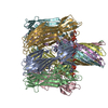
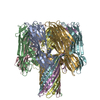
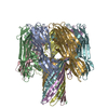
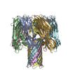
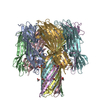
| Entry | Database: PDB / ID: 7ahl | ||||||
|---|---|---|---|---|---|---|---|
| Title | ALPHA-HEMOLYSIN FROM STAPHYLOCOCCUS AUREUS | ||||||
 Components Components | ALPHA-HEMOLYSIN | ||||||
 Keywords Keywords | CYTOLYTIC PROTEIN / HEMOLYSIN / TRANSMEMBRANE PORE | ||||||
| Function / homology |  Function and homology information Function and homology informationcytolysis in another organism / The NLRP3 inflammasome / Purinergic signaling in leishmaniasis infection / toxin activity / extracellular region / identical protein binding Similarity search - Function | ||||||
| Biological species |   Staphylococcus aureus (bacteria) Staphylococcus aureus (bacteria) | ||||||
| Method |  X-RAY DIFFRACTION / MIR / Resolution: 1.89 Å X-RAY DIFFRACTION / MIR / Resolution: 1.89 Å | ||||||
 Authors Authors | Song, L. / Hobaugh, M. / Shustak, C. / Cheley, S. / Bayley, H. / Gouaux, J.E. | ||||||
 Citation Citation | Journal: Science / Year: 1996 Title: Structure of staphylococcal alpha-hemolysin, a heptameric transmembrane pore. Authors: Song, L. / Hobaugh, M.R. / Shustak, C. / Cheley, S. / Bayley, H. / Gouaux, J.E. | ||||||
| History |
|
- Structure visualization
| Structure viewer | Molecule: MolmilJmol/JSmol |
|---|
- Downloads & links
-Download
| PDBx/mmCIF format | 7ahl.cif.gz | 522.1 KB | Display | PDBx/mmCIF format |
|---|---|---|---|---|
| PDB format | pdb7ahl.ent.gz | 437.9 KB | Display | PDB format |
| PDBx/mmJSON format | 7ahl.json.gz | Tree view | PDBx/mmJSON format | |
| Others |  Other downloads Other downloads |
-Validation report
| Arichive directory | https://data.pdbj.org/pub/pdb/validation_reports/ah/7ahlftp://data.pdbj.org/pub/pdb/validation_reports/ah/7ahl | HTTPS FTP |
|---|
-Related structure data
| Similar structure data |
|---|


-Links
 PDBj
PDBj


- Assembly
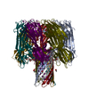
| Deposited unit | 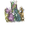
| ||||||||||||||||||||||||||||
|---|---|---|---|---|---|---|---|---|---|---|---|---|---|---|---|---|---|---|---|---|---|---|---|---|---|---|---|---|---|
| 1 |
| ||||||||||||||||||||||||||||
| Unit cell |
| ||||||||||||||||||||||||||||
| Noncrystallographic symmetry (NCS) | NCS oper:
|
-Components
| #1: Protein | Mass: 33290.000 Da / Num. of mol.: 7 / Source method: isolated from a natural source / Source: (natural) Staphylococcus aureus (bacteria) / Strain: WOOD46 / References: UniProt: P09616#2: Water | ChemComp-HOH / |  Mass: 18.015 Da / Num. of mol.: 818 / Source method: isolated from a natural source / Formula: H2O Mass: 18.015 Da / Num. of mol.: 818 / Source method: isolated from a natural source / Formula: H2O |
|---|
-Experimental details
-Experiment
| Experiment | Method: X-RAY DIFFRACTION / Number of used crystals: 3 |
|---|
- Sample preparation
| Crystal | Density Matthews: 3 Å3/Da / Density % sol: 60 % | ||||||||||||||||
|---|---|---|---|---|---|---|---|---|---|---|---|---|---|---|---|---|---|
| Crystal grow | pH: 6 Details: PROTEIN WAS CRYSTALLIZED BY MIXING EQUAL VOLUMES OF A PROTEIN SOLUTION OF 8 MG/ML PROTEIN, 25 MM BETA-OG, 10 MM TRIS AT PH 8.5 AND A PRECIPITANT SOLUTION OF 2.5 M (NH4)2SO4, 0.25% PEG 5K ...Details: PROTEIN WAS CRYSTALLIZED BY MIXING EQUAL VOLUMES OF A PROTEIN SOLUTION OF 8 MG/ML PROTEIN, 25 MM BETA-OG, 10 MM TRIS AT PH 8.5 AND A PRECIPITANT SOLUTION OF 2.5 M (NH4)2SO4, 0.25% PEG 5K MONOMETHYL ETHER AND 75MM SODIUM CACODYLATE, PH 6.0. | ||||||||||||||||
| Crystal grow | *PLUS Method: unknown | ||||||||||||||||
| Components of the solutions | *PLUS
|
-Data collection
| Diffraction | Mean temperature: 287 K |
|---|---|
| Diffraction source | Source: ROTATING ANODE / Type: RIGAKU RUH2R / Wavelength: 1.5418 |
| Detector | Type: RIGAKU / Detector: IMAGE PLATE / Date: Aug 1, 1994 / Details: MIRRORS |
| Radiation | Monochromator: NI FILTER / Monochromatic (M) / Laue (L): M / Scattering type: x-ray |
| Radiation wavelength | Wavelength: 1.5418 Å / Relative weight: 1 |
| Reflection | Resolution: 1.89→15 Å / Num. obs: 206863 / % possible obs: 93.3 % / Observed criterion σ(I): 2 / Redundancy: 14.1 % / Rmerge(I) obs: 0.08 / Rsym value: 0.08 / Net I/σ(I): 8.7 |
| Reflection shell | Resolution: 1.89→2 Å / Redundancy: 2 % / Rmerge(I) obs: 0.083 / Mean I/σ(I) obs: 3 / Rsym value: 0.349 / % possible all: 94.4 |
| Reflection | *PLUS Num. measured all: 2924665 |
| Reflection shell | *PLUS % possible obs: 94.4 % |
- Processing
| Software |
| ||||||||||||||||||||||||||||||||||||||||||||||||||||||||||||
|---|---|---|---|---|---|---|---|---|---|---|---|---|---|---|---|---|---|---|---|---|---|---|---|---|---|---|---|---|---|---|---|---|---|---|---|---|---|---|---|---|---|---|---|---|---|---|---|---|---|---|---|---|---|---|---|---|---|---|---|---|---|
| Refinement | Method to determine structure: MIR / Resolution: 1.89→8 Å / σ(F): 2
| ||||||||||||||||||||||||||||||||||||||||||||||||||||||||||||
| Refinement step | Cycle: LAST / Resolution: 1.89→8 Å
| ||||||||||||||||||||||||||||||||||||||||||||||||||||||||||||
| Refine LS restraints |
| ||||||||||||||||||||||||||||||||||||||||||||||||||||||||||||
| Software | *PLUS Name: X-PLOR / Version: 3.1 / Classification: refinement | ||||||||||||||||||||||||||||||||||||||||||||||||||||||||||||
| Refinement | *PLUS Num. reflection obs: 198341 | ||||||||||||||||||||||||||||||||||||||||||||||||||||||||||||
| Solvent computation | *PLUS | ||||||||||||||||||||||||||||||||||||||||||||||||||||||||||||
| Displacement parameters | *PLUS |
