protein localization to astral microtubule / cell cortex region / protein localization to mitotic spindle / neuron to neuron synapse / cortical microtubule cytoskeleton / mitotic spindle astral microtubule end / centriolar subdistal appendage / centriole-centriole cohesion / positive regulation of neuromuscular junction development / microtubule anchoring at centrosome ...protein localization to astral microtubule / cell cortex region / protein localization to mitotic spindle / neuron to neuron synapse / cortical microtubule cytoskeleton / mitotic spindle astral microtubule end / centriolar subdistal appendage / centriole-centriole cohesion / positive regulation of neuromuscular junction development / microtubule anchoring at centrosome / maintenance of synapse structure / ventral spinal cord development / protein localization to microtubule / mitotic nuclear membrane disassembly / dynein complex / cell projection membrane / microtubule plus-end / mitotic spindle microtubule / positive regulation of microtubule nucleation / XBP1(S) activates chaperone genes / attachment of mitotic spindle microtubules to kinetochore / positive regulation of dendritic spine morphogenesis / melanosome transport / microtubule plus-end binding / microtubule bundle formation / non-motile cilium assembly / protein localization to centrosome / retrograde transport, endosome to Golgi / COPI-independent Golgi-to-ER retrograde traffic / microtubule associated complex / nuclear migration / perinuclear theca / neuromuscular process / mitotic spindle pole / spindle midzone / negative regulation of microtubule polymerization / establishment of mitotic spindle orientation / neuromuscular junction development / motor behavior / cell leading edge / microtubule polymerization / microtubule organizing center / regulation of microtubule polymerization / regulation of microtubule polymerization or depolymerization / intercellular bridge / positive regulation of microtubule polymerization / COPI-mediated anterograde transport / cytoplasmic microtubule / spindle assembly / behavioral response to cocaine / neuron projection maintenance / Amplification of signal from unattached kinetochores via a MAD2 inhibitory signal / Loss of Nlp from mitotic centrosomes / Loss of proteins required for interphase microtubule organization from the centrosome / Recruitment of mitotic centrosome proteins and complexes / MHC class II antigen presentation / Recruitment of NuMA to mitotic centrosomes / Anchoring of the basal body to the plasma membrane / Mitotic Prometaphase / HSP90 chaperone cycle for steroid hormone receptors (SHR) in the presence of ligand / EML4 and NUDC in mitotic spindle formation / acrosomal vesicle / AURKA Activation by TPX2 / Resolution of Sister Chromatid Cohesion / regulation of mitotic spindle organization / protein serine/threonine kinase activator activity / protein serine/threonine kinase binding / centriole / tubulin binding / neuron cellular homeostasis / RHO GTPases Activate Formins / kinetochore / tau protein binding / spindle / spindle pole / mitotic spindle / The role of GTSE1 in G2/M progression after G2 checkpoint / Signaling by ALK fusions and activated point mutants / Separation of Sister Chromatids / intracellular protein localization / Regulation of PLK1 Activity at G2/M Transition / nuclear envelope / nervous system development / cell migration / mitotic cell cycle / microtubule cytoskeleton / midbody / cell cortex / microtubule binding / microtubule / neuron projection / cilium / ciliary basal body / cadherin binding / axon / cell division / focal adhesion / neuronal cell body / centrosome / protein kinase binding Similarity search - Function
Single alpha-helices involved in coiled-coils or other helix-helix interfaces - #1430 / CAP Gly-rich-like domain / Dynein associated protein / Dynein associated protein / EB1, C-terminal / Microtubule-associated protein RP/EB / EB1, C-terminal domain superfamily / EB1-C terminal (EB1-C) domain profile. / EB1-like C-terminal motif / CAP-Gly domain signature. ...Single alpha-helices involved in coiled-coils or other helix-helix interfaces - #1430 / CAP Gly-rich-like domain / Dynein associated protein / Dynein associated protein / EB1, C-terminal / Microtubule-associated protein RP/EB / EB1, C-terminal domain superfamily / EB1-C terminal (EB1-C) domain profile. / EB1-like C-terminal motif / CAP-Gly domain signature. / CAP Gly-rich domain / CAP Gly-rich domain superfamily / CAP-Gly domain / CAP-Gly domain profile. / CAP_GLY / Calponin homology (CH) domain / Calponin homology domain / CH domain superfamily / Calponin homology (CH) domain profile. / Single alpha-helices involved in coiled-coils or other helix-helix interfaces / SH3 type barrels. / Roll / Up-down Bundle / Mainly Beta / Mainly Alpha Similarity search - Domain/homology
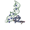
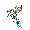
Dynactin subunit 1 / Microtubule-associated protein RP/EB family member 1 / Microtubule-associated protein RP/EB family member 3 Similarity search - Component
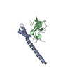
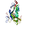
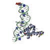
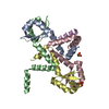
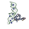
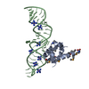
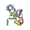
A: Microtubule-associated protein RP/EB family member 1 B: Microtubule-associated protein RP/EB family member 3 P: Dynactin subunit 1 Q: Dynactin subunit 1
In the structure databanks used in Yorodumi, some data are registered as the other names, "COVID-19 virus" and "2019-nCoV". Here are the details of the virus and the list of structure data.
Jan 31, 2019. EMDB accession codes are about to change! (news from PDBe EMDB page)
EMDB accession codes are about to change! (news from PDBe EMDB page)
The allocation of 4 digits for EMDB accession codes will soon come to an end. Whilst these codes will remain in use, new EMDB accession codes will include an additional digit and will expand incrementally as the available range of codes is exhausted. The current 4-digit format prefixed with “EMD-” (i.e. EMD-XXXX) will advance to a 5-digit format (i.e. EMD-XXXXX), and so on. It is currently estimated that the 4-digit codes will be depleted around Spring 2019, at which point the 5-digit format will come into force.
The EM Navigator/Yorodumi systems omit the EMD- prefix.
Related info.:Q: What is EMD? / ID/Accession-code notation in Yorodumi/EM Navigator
Yorodumi is a browser for structure data from EMDB, PDB, SASBDB, etc.
This page is also the successor to EM Navigator detail page, and also detail information page/front-end page for Omokage search.
The word "yorodu" (or yorozu) is an old Japanese word meaning "ten thousand". "mi" (miru) is to see.
Related info.:EMDB / PDB / SASBDB / Comparison of 3 databanks / Yorodumi Search / Aug 31, 2016. New EM Navigator & Yorodumi / Yorodumi Papers / Jmol/JSmol / Function and homology information / Changes in new EM Navigator and Yorodumi
 Movie
Movie Controller

 Yorodumi
 Open data
 Basic information
 Components
 Keywords
 Function and homology information
 Homo sapiens (human)
 X-RAY DIFFRACTION /
  Authors
 Citation
 Structure visualization
 Downloads & links
 Other downloads
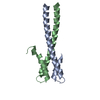


 PDBj
PDBj


 Assembly
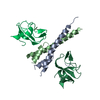

 Mass: 18.015 Da / Num. of mol.: 36 / Source method: isolated from a natural source / Formula: H2O
 Sample preparation
 / Beamline: X06SA / Wavelength: 1 Å
 Processing