 Movie
Movie Controller

[English] 日本語
 Yorodumi
- PDB-3to7: Crystal structure of yeast Esa1 HAT domain bound to coenzyme A wi... -
+ Open data

- Basic information
| Entry | Database: PDB / ID: 3to7 | ||||||
|---|---|---|---|---|---|---|---|
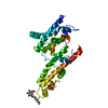
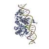
| Title | Crystal structure of yeast Esa1 HAT domain bound to coenzyme A with active site lysine acetylated | ||||||
 Components Components | Histone acetyltransferase ESA1 | ||||||
 Keywords Keywords | TRANSFERASE / MYST family / histone acetyltransferase | ||||||
| Function / homology |  Function and homology information Function and homology informationDNA Damage/Telomere Stress Induced Senescence / Sensing of DNA Double Strand Breaks / piccolo histone acetyltransferase complex / peptide 2-hydroxyisobutyryltransferase activity / histone crotonyltransferase activity / positive regulation of triglyceride biosynthetic process / histone H4 acetyltransferase activity / rDNA heterochromatin formation / DNA-templated transcription elongation / Recruitment and ATM-mediated phosphorylation of repair and signaling proteins at DNA double strand breaks ...DNA Damage/Telomere Stress Induced Senescence / Sensing of DNA Double Strand Breaks / piccolo histone acetyltransferase complex / peptide 2-hydroxyisobutyryltransferase activity / histone crotonyltransferase activity / positive regulation of triglyceride biosynthetic process / histone H4 acetyltransferase activity / rDNA heterochromatin formation / DNA-templated transcription elongation / Recruitment and ATM-mediated phosphorylation of repair and signaling proteins at DNA double strand breaks / histone acetyltransferase activity / NuA4 histone acetyltransferase complex / Estrogen-dependent gene expression / positive regulation of macroautophagy / protein-lysine-acetyltransferase activity / histone acetyltransferase / Transferases; Acyltransferases; Transferring groups other than aminoacyl groups / transcription coregulator activity / positive regulation of transcription elongation by RNA polymerase II / nucleosome / regulation of cell cycle / DNA repair / chromatin binding / regulation of transcription by RNA polymerase II / chromatin / DNA-templated transcription / nucleus Similarity search - Function | ||||||
| Biological species |  | ||||||
| Method |  X-RAY DIFFRACTION / SYNCHROTRON / MOLECULAR REPLACEMENT / Resolution: 1.9 Å X-RAY DIFFRACTION / SYNCHROTRON / MOLECULAR REPLACEMENT / Resolution: 1.9 Å | ||||||
 Authors Authors | Yuan, H. / Ding, E.C. / Marmorstein, R. | ||||||
 Citation Citation | Journal: Embo J. / Year: 2011 Title: MYST protein acetyltransferase activity requires active site lysine autoacetylation. Authors: Yuan, H. / Rossetto, D. / Mellert, H. / Dang, W. / Srinivasan, M. / Johnson, J. / Hodawadekar, S. / Ding, E.C. / Speicher, K. / Abshiru, N. / Perry, R. / Wu, J. / Yang, C. / Zheng, Y.G. / ...Authors: Yuan, H. / Rossetto, D. / Mellert, H. / Dang, W. / Srinivasan, M. / Johnson, J. / Hodawadekar, S. / Ding, E.C. / Speicher, K. / Abshiru, N. / Perry, R. / Wu, J. / Yang, C. / Zheng, Y.G. / Speicher, D.W. / Thibault, P. / Verreault, A. / Johnson, F.B. / Berger, S.L. / Sternglanz, R. / McMahon, S.B. / Cote, J. / Marmorstein, R. | ||||||
| History |
|
- Structure visualization
| Structure viewer | Molecule: MolmilJmol/JSmol |
|---|
- Downloads & links
-Download
| PDBx/mmCIF format | 3to7.cif.gz | 79.5 KB | Display | PDBx/mmCIF format |
|---|---|---|---|---|
| PDB format | pdb3to7.ent.gz | 58.8 KB | Display | PDB format |
| PDBx/mmJSON format | 3to7.json.gz | Tree view | PDBx/mmJSON format | |
| Others |  Other downloads Other downloads |
-Validation report
| Arichive directory | https://data.pdbj.org/pub/pdb/validation_reports/to/3to7ftp://data.pdbj.org/pub/pdb/validation_reports/to/3to7 | HTTPS FTP |
|---|
-Related structure data
| Related structure data |  3to6C  3to9C  3toaC  3tobC 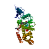 1mjaS C: citing same article ( S: Starting model for refinement |
|---|---|
| Similar structure data |


-Links
 PDBj
PDBj


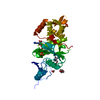
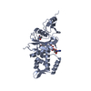
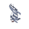
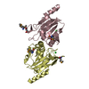
- Assembly
| Deposited unit | 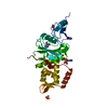
| ||||||||
|---|---|---|---|---|---|---|---|---|---|
| 1 |
| ||||||||
| 2 | 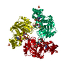
| ||||||||
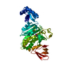
| Unit cell |
|
-Components
-Protein , 1 types, 1 molecules A
| #1: Protein | Mass: 33153.070 Da / Num. of mol.: 1 / Fragment: UNP residues 160-435 Source method: isolated from a genetically manipulated source Source: (gene. exp.) Gene: ESA1, YOR244W, O5257 / Production host:  |
|---|
-Non-polymers , 5 types, 198 molecules 
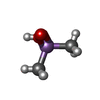


| #2: Chemical | ChemComp-COA /  Mass: 767.534 Da / Num. of mol.: 1 / Source method: obtained synthetically / Formula: C21H36N7O16P3S Mass: 767.534 Da / Num. of mol.: 1 / Source method: obtained synthetically / Formula: C21H36N7O16P3S | ||||
|---|---|---|---|---|---|
| #3: Chemical | ChemComp-CAD /  Mass: 137.997 Da / Num. of mol.: 1 / Source method: obtained synthetically / Formula: C2H7AsO2 Mass: 137.997 Da / Num. of mol.: 1 / Source method: obtained synthetically / Formula: C2H7AsO2 | ||||
| #4: Chemical |  Mass: 92.094 Da / Num. of mol.: 2 / Source method: obtained synthetically / Formula: C3H8O3 Mass: 92.094 Da / Num. of mol.: 2 / Source method: obtained synthetically / Formula: C3H8O3#5: Chemical | ChemComp-SO4 / |  Mass: 96.063 Da / Num. of mol.: 1 / Source method: obtained synthetically / Formula: SO4 Mass: 96.063 Da / Num. of mol.: 1 / Source method: obtained synthetically / Formula: SO4#6: Water | ChemComp-HOH / | Mass: 18.015 Da / Num. of mol.: 193 / Source method: isolated from a natural source / Formula: H2O |
-Details
| Has protein modification | Y |
|---|
-Experimental details
-Experiment
| Experiment | Method: X-RAY DIFFRACTION / Number of used crystals: 1 |
|---|
- Sample preparation
| Crystal | Density Matthews: 3.85 Å3/Da / Density % sol: 68.08 % |
|---|---|
| Crystal grow | Temperature: 298 K / Method: vapor diffusion, hanging drop / pH: 6.5 Details: 0.1 M sodium cacodylate, 1.6 M ammonium sulfate, pH 6.5, VAPOR DIFFUSION, HANGING DROP, temperature 298.0K |
-Data collection
| Diffraction | Mean temperature: 100 K |
|---|---|
| Diffraction source | Source: SYNCHROTRON / Site: NSLS  / Beamline: X6A / Wavelength: 1.127 Å / Beamline: X6A / Wavelength: 1.127 Å |
| Detector | Type: ADSC QUANTUM 270 / Detector: CCD / Date: Feb 11, 2011 / Details: TOROIDAL FOCUSING MIRROR |
| Radiation | Protocol: SINGLE WAVELENGTH / Monochromatic (M) / Laue (L): M / Scattering type: x-ray |
| Radiation wavelength | Wavelength: 1.127 Å / Relative weight: 1 |
| Reflection | Resolution: 1.9→50 Å / Num. all: 41363 / Num. obs: 41363 / % possible obs: 100 % / Observed criterion σ(F): 0 / Observed criterion σ(I): 0 / Redundancy: 10.8 % / Rmerge(I) obs: 0.088 / Net I/σ(I): 27.2 |
| Reflection shell | Resolution: 1.9→1.93 Å / Redundancy: 10.8 % / Rmerge(I) obs: 0.416 / Mean I/σ(I) obs: 6.4 / Num. unique all: 2045 / % possible all: 100 |
- Processing
| Software |
| ||||||||||||||||||||||||||||||||||||||||||||||||||||||||||||||||||||||||||||||||||||||||||||||||||||||||||||||||
|---|---|---|---|---|---|---|---|---|---|---|---|---|---|---|---|---|---|---|---|---|---|---|---|---|---|---|---|---|---|---|---|---|---|---|---|---|---|---|---|---|---|---|---|---|---|---|---|---|---|---|---|---|---|---|---|---|---|---|---|---|---|---|---|---|---|---|---|---|---|---|---|---|---|---|---|---|---|---|---|---|---|---|---|---|---|---|---|---|---|---|---|---|---|---|---|---|---|---|---|---|---|---|---|---|---|---|---|---|---|---|---|---|---|
| Refinement | Method to determine structure: MOLECULAR REPLACEMENT Starting model: PDB ENTRY 1MJA Resolution: 1.9→30.507 Å / SU ML: 0.22 / σ(F): 0.03 / Phase error: 21.19 / Stereochemistry target values: ML
| ||||||||||||||||||||||||||||||||||||||||||||||||||||||||||||||||||||||||||||||||||||||||||||||||||||||||||||||||
| Solvent computation | Shrinkage radii: 0.83 Å / VDW probe radii: 1.1 Å / Solvent model: FLAT BULK SOLVENT MODEL / Bsol: 40 Å2 / ksol: 0.4 e/Å3 | ||||||||||||||||||||||||||||||||||||||||||||||||||||||||||||||||||||||||||||||||||||||||||||||||||||||||||||||||
| Displacement parameters | Biso mean: 19.18 Å2
| ||||||||||||||||||||||||||||||||||||||||||||||||||||||||||||||||||||||||||||||||||||||||||||||||||||||||||||||||
| Refinement step | Cycle: LAST / Resolution: 1.9→30.507 Å
| ||||||||||||||||||||||||||||||||||||||||||||||||||||||||||||||||||||||||||||||||||||||||||||||||||||||||||||||||
| Refine LS restraints |
| ||||||||||||||||||||||||||||||||||||||||||||||||||||||||||||||||||||||||||||||||||||||||||||||||||||||||||||||||
| LS refinement shell |
|
