 Movie
Movie Controller


+ Open data

- Basic information
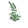
| Entry | Database: PDB / ID: 3keb | ||||||
|---|---|---|---|---|---|---|---|
| Title | Thiol peroxidase from Chromobacterium violaceum | ||||||
 Components Components | Probable thiol peroxidase | ||||||
 Keywords Keywords | OXIDOREDUCTASE / structural genomics / APC40679 / thiol peroxidase. / PSI-2 / Protein Structure Initiative / Midwest Center for Structural Genomics / MCSG / Peroxidase | ||||||
| Function / homology |  Function and homology information Function and homology informationOxidoreductases; Acting on a peroxide as acceptor; Peroxidases / peroxidase activity Similarity search - Function | ||||||
| Biological species |  Chromobacterium violaceum (bacteria) Chromobacterium violaceum (bacteria) | ||||||
| Method |  X-RAY DIFFRACTION / SYNCHROTRON / MOLECULAR REPLACEMENT / Resolution: 1.8 Å X-RAY DIFFRACTION / SYNCHROTRON / MOLECULAR REPLACEMENT / Resolution: 1.8 Å | ||||||
 Authors Authors | Osipiuk, J. / Kagan, O. / Savchenko, A. / Edwards, A.M. / Joachimiak, A. / Midwest Center for Structural Genomics (MCSG) | ||||||
 Citation Citation | Journal: To be Published Title: X-ray crystal structure of thiol peroxidase from Chromobacterium violaceum Authors: Osipiuk, J. / Kagan, O. / Savchenko, A. / Edwards, A.M. / Joachimiak, A. | ||||||
| History |
|
- Structure visualization
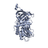
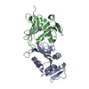
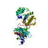
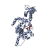
| Structure viewer | Molecule: MolmilJmol/JSmol |
|---|
- Downloads & links
-Download
| PDBx/mmCIF format | 3keb.cif.gz | 173.9 KB | Display | PDBx/mmCIF format |
|---|---|---|---|---|
| PDB format | pdb3keb.ent.gz | 139.1 KB | Display | PDB format |
| PDBx/mmJSON format | 3keb.json.gz | Tree view | PDBx/mmJSON format | |
| Others |  Other downloads Other downloads |
-Validation report
| Arichive directory | https://data.pdbj.org/pub/pdb/validation_reports/ke/3kebftp://data.pdbj.org/pub/pdb/validation_reports/ke/3keb | HTTPS FTP |
|---|
-Related structure data
| Related structure data |  1q98S S: Starting model for refinement |
|---|---|
| Similar structure data | |
| Other databases |


-Links
 PDBj
PDBj- Assembly
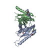
| Deposited unit | 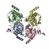
| ||||||||
|---|---|---|---|---|---|---|---|---|---|
| 1 | 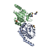
| ||||||||
| 2 | 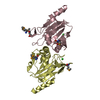
| ||||||||
| Unit cell |
|
-Components
| #1: Protein | Mass: 25615.123 Da / Num. of mol.: 4 Source method: isolated from a genetically manipulated source Source: (gene. exp.) Chromobacterium violaceum (bacteria) / Strain: ATCC 12472 / Gene: CV_3708 / Plasmid: pET15b modified / Production host: References: UniProt: Q7NRS2, Oxidoreductases; Acting on a peroxide as acceptor; Peroxidases #2: Chemical |   Mass: 96.063 Da / Num. of mol.: 2 / Source method: obtained synthetically / Formula: SO4 Mass: 96.063 Da / Num. of mol.: 2 / Source method: obtained synthetically / Formula: SO4#3: Chemical |   Mass: 35.453 Da / Num. of mol.: 3 / Source method: obtained synthetically / Formula: Cl Mass: 35.453 Da / Num. of mol.: 3 / Source method: obtained synthetically / Formula: Cl#4: Water | ChemComp-HOH / |  Mass: 18.015 Da / Num. of mol.: 761 / Source method: isolated from a natural source / Formula: H2O Mass: 18.015 Da / Num. of mol.: 761 / Source method: isolated from a natural source / Formula: H2OHas protein modification | Y | |
|---|
-Experimental details
-Experiment
| Experiment | Method: X-RAY DIFFRACTION / Number of used crystals: 1 |
|---|
- Sample preparation
| Crystal | Density Matthews: 2 Å3/Da / Density % sol: 38.52 % |
|---|---|
| Crystal grow | Temperature: 294 K / Method: vapor diffusion, hanging drop / pH: 7.5 Details: 20% PEG-3350, 0.2 M ammonium sulfate, pH 7.5, VAPOR DIFFUSION, HANGING DROP, temperature 294K |
-Data collection
| Diffraction | Mean temperature: 100 K |
|---|---|
| Diffraction source | Source: SYNCHROTRON / Site: APS  / Beamline: 19-ID / Wavelength: 0.9792 Å / Beamline: 19-ID / Wavelength: 0.9792 Å |
| Detector | Type: ADSC QUANTUM 315 / Detector: CCD / Date: Oct 18, 2009 |
| Radiation | Monochromator: double crystal monochromator / Protocol: SINGLE WAVELENGTH / Monochromatic (M) / Laue (L): M / Scattering type: x-ray |
| Radiation wavelength | Wavelength: 0.9792 Å / Relative weight: 1 |
| Reflection | Resolution: 1.8→43 Å / Num. all: 74165 / Num. obs: 74165 / % possible obs: 99 % / Observed criterion σ(F): 0 / Observed criterion σ(I): 0 / Redundancy: 4 % / Biso Wilson estimate: 29.2 Å2 / Rmerge(I) obs: 0.087 / Χ2: 2.218 / Net I/σ(I): 8.2 |
| Reflection shell | Resolution: 1.8→1.83 Å / Redundancy: 2.9 % / Rmerge(I) obs: 0.502 / Mean I/σ(I) obs: 2.13 / Num. unique all: 3250 / Χ2: 1.292 / % possible all: 88.5 |
- Processing
| Software |
| |||||||||||||||||||||||||||||||||||||||||||||||||||||||||||||||||||||||||||||||||||||||||||||||||||||||||||||||||||||||||||||
|---|---|---|---|---|---|---|---|---|---|---|---|---|---|---|---|---|---|---|---|---|---|---|---|---|---|---|---|---|---|---|---|---|---|---|---|---|---|---|---|---|---|---|---|---|---|---|---|---|---|---|---|---|---|---|---|---|---|---|---|---|---|---|---|---|---|---|---|---|---|---|---|---|---|---|---|---|---|---|---|---|---|---|---|---|---|---|---|---|---|---|---|---|---|---|---|---|---|---|---|---|---|---|---|---|---|---|---|---|---|---|---|---|---|---|---|---|---|---|---|---|---|---|---|---|---|---|
| Refinement | Method to determine structure: MOLECULAR REPLACEMENT Starting model: PDB entry 1Q98 Resolution: 1.8→43 Å / Cor.coef. Fo:Fc: 0.977 / Cor.coef. Fo:Fc free: 0.962 / Occupancy max: 1 / Occupancy min: 0.24 / SU B: 6.975 / SU ML: 0.094 / TLS residual ADP flag: LIKELY RESIDUAL / Cross valid method: THROUGHOUT / σ(F): 0 / σ(I): 0 / ESU R: 0.148 / ESU R Free: 0.137 / Stereochemistry target values: MAXIMUM LIKELIHOOD Details: HYDROGENS HAVE BEEN ADDED IN THE RIDING POSITIONS, U VALUES RESIDUAL ONLY
| |||||||||||||||||||||||||||||||||||||||||||||||||||||||||||||||||||||||||||||||||||||||||||||||||||||||||||||||||||||||||||||
| Solvent computation | Ion probe radii: 0.8 Å / Shrinkage radii: 0.8 Å / VDW probe radii: 1.4 Å / Solvent model: MASK | |||||||||||||||||||||||||||||||||||||||||||||||||||||||||||||||||||||||||||||||||||||||||||||||||||||||||||||||||||||||||||||
| Displacement parameters | Biso max: 54.17 Å2 / Biso mean: 18.685 Å2 / Biso min: 2.95 Å2
| |||||||||||||||||||||||||||||||||||||||||||||||||||||||||||||||||||||||||||||||||||||||||||||||||||||||||||||||||||||||||||||
| Refinement step | Cycle: LAST / Resolution: 1.8→43 Å
| |||||||||||||||||||||||||||||||||||||||||||||||||||||||||||||||||||||||||||||||||||||||||||||||||||||||||||||||||||||||||||||
| Refine LS restraints |
| |||||||||||||||||||||||||||||||||||||||||||||||||||||||||||||||||||||||||||||||||||||||||||||||||||||||||||||||||||||||||||||
| LS refinement shell | Resolution: 1.799→1.846 Å / Total num. of bins used: 20
| |||||||||||||||||||||||||||||||||||||||||||||||||||||||||||||||||||||||||||||||||||||||||||||||||||||||||||||||||||||||||||||
| Refinement TLS params. | Method: refined / Refine-ID: X-RAY DIFFRACTION
| |||||||||||||||||||||||||||||||||||||||||||||||||||||||||||||||||||||||||||||||||||||||||||||||||||||||||||||||||||||||||||||
| Refinement TLS group |
|
