 Movie
Movie Controller

[English] 日本語
 Yorodumi
- PDB-3fg9: The crystal structure of an universal stress protein UspA family ... -
+ Open data

- Basic information
| Entry | Database: PDB / ID: 3fg9 | ||||||
|---|---|---|---|---|---|---|---|
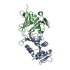
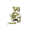
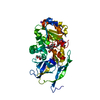
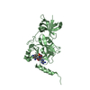
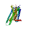
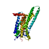
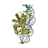
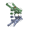
| Title | The crystal structure of an universal stress protein UspA family protein from Lactobacillus plantarum WCFS1 | ||||||
 Components Components | protein of universal stress protein UspA family | ||||||
 Keywords Keywords | structural genomics / unknown function / APC60691 / universal stress protein UspA family / nucleotide-binding / Lactobacillus plantarum WCFS1 / PSI-2 / protein structure initiative / midwest center for structural genomics / MCSG | ||||||
| Function / homology |  Function and homology information Function and homology information | ||||||
| Biological species |  Lactobacillus plantarum (bacteria) Lactobacillus plantarum (bacteria) | ||||||
| Method |  X-RAY DIFFRACTION / SYNCHROTRON / SAD / Resolution: 1.47 Å X-RAY DIFFRACTION / SYNCHROTRON / SAD / Resolution: 1.47 Å | ||||||
 Authors Authors | Tan, K. / Li, H. / Cobb, G. / Joachimiak, A. / Midwest Center for Structural Genomics (MCSG) | ||||||
 Citation Citation | Journal: To be Published Title: The crystal structure of an universal stress protein UspA family protein from Lactobacillus plantarum WCFS1 Authors: Tan, K. / Li, H. / Cobb, G. / Joachimiak, A. | ||||||
| History |
|
- Structure visualization
| Structure viewer | Molecule: MolmilJmol/JSmol |
|---|
- Downloads & links
-Download
| PDBx/mmCIF format | 3fg9.cif.gz | 397.1 KB | Display | PDBx/mmCIF format |
|---|---|---|---|---|
| PDB format | pdb3fg9.ent.gz | 326.6 KB | Display | PDB format |
| PDBx/mmJSON format | 3fg9.json.gz | Tree view | PDBx/mmJSON format | |
| Others |  Other downloads Other downloads |
-Validation report
| Arichive directory | https://data.pdbj.org/pub/pdb/validation_reports/fg/3fg9ftp://data.pdbj.org/pub/pdb/validation_reports/fg/3fg9 | HTTPS FTP |
|---|
-Related structure data
| Similar structure data | |
|---|---|
| Other databases |


-Links
 PDBj
PDBj- Assembly
| Deposited unit | 
| ||||||||
|---|---|---|---|---|---|---|---|---|---|
| 1 | 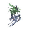
| ||||||||
| 2 | 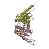
| ||||||||
| 3 | 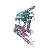
| ||||||||
| Unit cell |
| ||||||||
| Details | Experimentally unknown. It is predicted that chains A and B, C and D, E and F form dimers respectively. |
-Components
| #1: Protein | Mass: 17504.408 Da / Num. of mol.: 6 Source method: isolated from a genetically manipulated source Source: (gene. exp.) Lactobacillus plantarum (bacteria) / Strain: WCFS1 / Gene: Lactobacillus plantarum, lp_3663 / Plasmid: pMCSG7 / Production host: #2: Chemical | ChemComp-MG /   Mass: 24.305 Da / Num. of mol.: 7 / Source method: obtained synthetically / Formula: Mg Mass: 24.305 Da / Num. of mol.: 7 / Source method: obtained synthetically / Formula: Mg#3: Chemical | ChemComp-FMT /   Mass: 46.025 Da / Num. of mol.: 7 / Source method: obtained synthetically / Formula: CH2O2 Mass: 46.025 Da / Num. of mol.: 7 / Source method: obtained synthetically / Formula: CH2O2#4: Chemical | ChemComp-ACT / |   Mass: 59.044 Da / Num. of mol.: 1 / Source method: obtained synthetically / Formula: C2H3O2 Mass: 59.044 Da / Num. of mol.: 1 / Source method: obtained synthetically / Formula: C2H3O2#5: Water | ChemComp-HOH / |  Mass: 18.015 Da / Num. of mol.: 943 / Source method: isolated from a natural source / Formula: H2O Mass: 18.015 Da / Num. of mol.: 943 / Source method: isolated from a natural source / Formula: H2OHas protein modification | Y | |
|---|
-Experimental details
-Experiment
| Experiment | Method: X-RAY DIFFRACTION / Number of used crystals: 1 |
|---|
- Sample preparation
| Crystal | Density Matthews: 2.08 Å3/Da / Density % sol: 41 % |
|---|---|
| Crystal grow | Temperature: 289 K / Method: vapor diffusion, sitting drop Details: 0.2M MgActate. 20% PEG3350, VAPOR DIFFUSION, SITTING DROP, temperature 289K |
-Data collection
| Diffraction | Mean temperature: 100 K |
|---|---|
| Diffraction source | Source: SYNCHROTRON / Site: APS  / Beamline: 19-ID / Wavelength: 0.97942 Å / Beamline: 19-ID / Wavelength: 0.97942 Å |
| Detector | Type: ADSC QUANTUM 315 / Detector: CCD / Date: Oct 8, 2008 / Details: Mirror |
| Radiation | Monochromator: Si 111 crystal / Protocol: SINGLE WAVELENGTH / Monochromatic (M) / Laue (L): M / Scattering type: x-ray |
| Radiation wavelength | Wavelength: 0.97942 Å / Relative weight: 1 |
| Reflection | Resolution: 1.47→37.6 Å / Num. all: 149737 / Num. obs: 149737 / % possible obs: 99.5 % / Observed criterion σ(F): 0 / Observed criterion σ(I): 0 / Redundancy: 8.4 % / Rmerge(I) obs: 0.068 / Net I/σ(I): 46.3 |
| Reflection shell | Resolution: 1.47→1.5 Å / Redundancy: 5.7 % / Rmerge(I) obs: 0.558 / Mean I/σ(I) obs: 2.6 / Num. unique all: 6828 / % possible all: 92.7 |
- Processing
| Software |
| ||||||||||||||||||||||||||||||||||||||||||||||||||||||||||||||||||||||||||||||||||||||||||||||||||||||||||||||||||||||||||||||||||||||||||||||||||||||||||||||||||||||||||
|---|---|---|---|---|---|---|---|---|---|---|---|---|---|---|---|---|---|---|---|---|---|---|---|---|---|---|---|---|---|---|---|---|---|---|---|---|---|---|---|---|---|---|---|---|---|---|---|---|---|---|---|---|---|---|---|---|---|---|---|---|---|---|---|---|---|---|---|---|---|---|---|---|---|---|---|---|---|---|---|---|---|---|---|---|---|---|---|---|---|---|---|---|---|---|---|---|---|---|---|---|---|---|---|---|---|---|---|---|---|---|---|---|---|---|---|---|---|---|---|---|---|---|---|---|---|---|---|---|---|---|---|---|---|---|---|---|---|---|---|---|---|---|---|---|---|---|---|---|---|---|---|---|---|---|---|---|---|---|---|---|---|---|---|---|---|---|---|---|---|---|---|
| Refinement | Method to determine structure: SAD / Resolution: 1.47→37.6 Å / Cor.coef. Fo:Fc: 0.963 / Cor.coef. Fo:Fc free: 0.951 / SU B: 2.301 / SU ML: 0.041 / Cross valid method: THROUGHOUT / σ(F): 0 / σ(I): 0 / ESU R: 0.092 / ESU R Free: 0.077 / Stereochemistry target values: MAXIMUM LIKELIHOOD / Details: HYDROGENS HAVE BEEN ADDED IN THE RIDING POSITIONS
| ||||||||||||||||||||||||||||||||||||||||||||||||||||||||||||||||||||||||||||||||||||||||||||||||||||||||||||||||||||||||||||||||||||||||||||||||||||||||||||||||||||||||||
| Solvent computation | Ion probe radii: 0.8 Å / Shrinkage radii: 0.8 Å / VDW probe radii: 1.2 Å / Solvent model: MASK | ||||||||||||||||||||||||||||||||||||||||||||||||||||||||||||||||||||||||||||||||||||||||||||||||||||||||||||||||||||||||||||||||||||||||||||||||||||||||||||||||||||||||||
| Displacement parameters | Biso mean: 20.385 Å2
| ||||||||||||||||||||||||||||||||||||||||||||||||||||||||||||||||||||||||||||||||||||||||||||||||||||||||||||||||||||||||||||||||||||||||||||||||||||||||||||||||||||||||||
| Refinement step | Cycle: LAST / Resolution: 1.47→37.6 Å
| ||||||||||||||||||||||||||||||||||||||||||||||||||||||||||||||||||||||||||||||||||||||||||||||||||||||||||||||||||||||||||||||||||||||||||||||||||||||||||||||||||||||||||
| Refine LS restraints |
| ||||||||||||||||||||||||||||||||||||||||||||||||||||||||||||||||||||||||||||||||||||||||||||||||||||||||||||||||||||||||||||||||||||||||||||||||||||||||||||||||||||||||||
| LS refinement shell | Resolution: 1.471→1.509 Å / Total num. of bins used: 20
|
