| Entry | Database: PDB / ID: 3tld
|
|---|
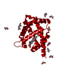
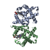
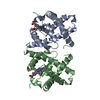
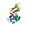
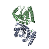
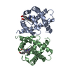
| Title | Crystal Structure of Y29F mutant of Vitreoscilla hemoglobin |
|---|
 Components Components | Bacterial hemoglobin |
|---|
 Keywords Keywords | OXYGEN TRANSPORT / globin 8-helix fold / oxygen storage |
|---|
| Function / homology |  Function and homology information Function and homology information
cellular response to nitrosative stress / nitric oxide dioxygenase [NAD(P)H] activity / nitric oxide catabolic process / FAD binding / oxygen carrier activity / oxygen binding / heme binding / metal ion bindingSimilarity search - Function Globin/Protoglobin / Globins / Globin-like / Globin / Globin / Globin domain profile. / Globin-like superfamily / Orthogonal Bundle / Mainly AlphaSimilarity search - Domain/homology |
|---|
| Biological species |  Vitreoscilla stercoraria (bacteria) Vitreoscilla stercoraria (bacteria) |
|---|
| Method |  X-RAY DIFFRACTION / SYNCHROTRON / MOLECULAR REPLACEMENT / Resolution: 1.896 Å X-RAY DIFFRACTION / SYNCHROTRON / MOLECULAR REPLACEMENT / Resolution: 1.896 Å |
|---|
 Authors Authors | Ratakonda, S. / Anand, A. / Dikshit, K. / Stark, B.C. / Howard, A.J. |
|---|
 Citation Citation | Journal: Acta Crystallogr.,Sect.F / Year: 2013
Title: Crystallographic structure determination of B10 mutants of Vitreoscilla hemoglobin: role of Tyr29 (B10) in the structure of the ligand-binding site.
Authors: Ratakonda, S. / Anand, A. / Dikshit, K. / Stark, B.C. / Howard, A.J. |
|---|
| History | | Deposition | Aug 29, 2011 | Deposition site: RCSB / Processing site: RCSB |
|---|
| Revision 1.0 | Apr 16, 2014 | Provider: repository / Type: Initial release |
|---|
| Revision 1.1 | Jul 17, 2019 | Group: Data collection / Refinement description / Category: software
Item: _software.classification / _software.name / _software.version |
|---|
| Revision 1.2 | Feb 28, 2024 | Group: Data collection / Database references / Derived calculations
Category: chem_comp_atom / chem_comp_bond ...chem_comp_atom / chem_comp_bond / database_2 / pdbx_struct_conn_angle / struct_conn / struct_ref_seq_dif / struct_site
Item: _database_2.pdbx_DOI / _database_2.pdbx_database_accession ..._database_2.pdbx_DOI / _database_2.pdbx_database_accession / _pdbx_struct_conn_angle.ptnr1_auth_asym_id / _pdbx_struct_conn_angle.ptnr1_label_asym_id / _pdbx_struct_conn_angle.ptnr2_auth_asym_id / _pdbx_struct_conn_angle.ptnr2_label_asym_id / _pdbx_struct_conn_angle.ptnr3_auth_asym_id / _pdbx_struct_conn_angle.ptnr3_label_asym_id / _pdbx_struct_conn_angle.value / _struct_conn.pdbx_dist_value / _struct_conn.ptnr1_auth_asym_id / _struct_conn.ptnr1_label_asym_id / _struct_conn.ptnr2_auth_asym_id / _struct_conn.ptnr2_label_asym_id / _struct_ref_seq_dif.details / _struct_site.pdbx_auth_asym_id / _struct_site.pdbx_auth_comp_id / _struct_site.pdbx_auth_seq_id |
|---|
|
|---|
 Movie
Movie Controller


 Open data
 Basic information
 Structure visualization
 Downloads & links
 Other downloads
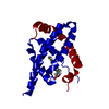


 PDBj
PDBj


 Assembly
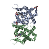

 Mass: 616.487 Da / Num. of mol.: 2 / Source method: obtained synthetically / Formula: C34H32FeN4O4
 Mass: 92.094 Da / Num. of mol.: 3 / Source method: obtained synthetically / Formula: C3H8O3
 Mass: 18.015 Da / Num. of mol.: 241 / Source method: isolated from a natural source / Formula: H2O
 Sample preparation
 / Beamline: 22-BM / Wavelength: 1 Å
 Processing