 Movie
Movie Controller

[English] 日本語
 Yorodumi
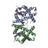
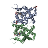
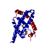
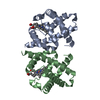
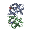
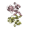
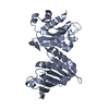
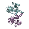
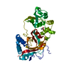
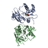
- PDB-2vhb: AZIDE ADDUCT OF THE BACTERIAL HEMOGLOBIN FROM VITREOSCILLA STERCORARIA -
+ Open data

- Basic information
| Entry | Database: PDB / ID: 2vhb | ||||||
|---|---|---|---|---|---|---|---|
| Title | AZIDE ADDUCT OF THE BACTERIAL HEMOGLOBIN FROM VITREOSCILLA STERCORARIA | ||||||
 Components Components | HEMOGLOBIN | ||||||
 Keywords Keywords | OXYGEN TRANSPORT / HEME / RESPIRATORY PROTEIN | ||||||
| Function / homology |  Function and homology information Function and homology informationcellular response to nitrosative stress / nitric oxide dioxygenase [NAD(P)H] activity / nitric oxide catabolic process / FAD binding / oxygen carrier activity / oxygen binding / heme binding / metal ion binding Similarity search - Function | ||||||
| Biological species |  Vitreoscilla stercoraria (bacteria) Vitreoscilla stercoraria (bacteria) | ||||||
| Method |  X-RAY DIFFRACTION / DIFFERENCE FOURIER / Resolution: 1.76 Å X-RAY DIFFRACTION / DIFFERENCE FOURIER / Resolution: 1.76 Å | ||||||
 Authors Authors | Tarricone, C. / Galizzi, A. / Coda, A. / Ascenzi, P. / Bolognesi, M. | ||||||
 Citation Citation | Journal: Structure / Year: 1997 Title: Unusual structure of the oxygen-binding site in the dimeric bacterial hemoglobin from Vitreoscilla sp. Authors: Tarricone, C. / Galizzi, A. / Coda, A. / Ascenzi, P. / Bolognesi, M. | ||||||
| History |
|
- Structure visualization
| Structure viewer | Molecule: MolmilJmol/JSmol |
|---|
- Downloads & links
-Download
| PDBx/mmCIF format | 2vhb.cif.gz | 70.2 KB | Display | PDBx/mmCIF format |
|---|---|---|---|---|
| PDB format | pdb2vhb.ent.gz | 52.5 KB | Display | PDB format |
| PDBx/mmJSON format | 2vhb.json.gz | Tree view | PDBx/mmJSON format | |
| Others |  Other downloads Other downloads |
-Validation report
| Arichive directory | https://data.pdbj.org/pub/pdb/validation_reports/vh/2vhbftp://data.pdbj.org/pub/pdb/validation_reports/vh/2vhb | HTTPS FTP |
|---|
-Related structure data

-Links
 PDBj
PDBj


- Assembly
| Deposited unit | 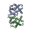
| ||||||||
|---|---|---|---|---|---|---|---|---|---|
| 1 |
| ||||||||
| Unit cell |
| ||||||||
| Noncrystallographic symmetry (NCS) | NCS oper: (Code: given Matrix: (0.24016, -0.00448, -0.97072), Vector: |
-Components
| #1: Protein | Mass: 15790.269 Da / Num. of mol.: 2 Source method: isolated from a genetically manipulated source Source: (gene. exp.) Vitreoscilla stercoraria (bacteria) / Strain: C1 / Gene: VGB / Plasmid: PDH88 / Gene (production host): VGB / Production host: #2: Chemical |   Mass: 42.020 Da / Num. of mol.: 2 / Source method: obtained synthetically / Formula: N3 Mass: 42.020 Da / Num. of mol.: 2 / Source method: obtained synthetically / Formula: N3#3: Chemical |   Mass: 616.487 Da / Num. of mol.: 2 / Source method: obtained synthetically / Formula: C34H32FeN4O4 Mass: 616.487 Da / Num. of mol.: 2 / Source method: obtained synthetically / Formula: C34H32FeN4O4#4: Water | ChemComp-HOH / |  Mass: 18.015 Da / Num. of mol.: 184 / Source method: isolated from a natural source / Formula: H2O Mass: 18.015 Da / Num. of mol.: 184 / Source method: isolated from a natural source / Formula: H2O |
|---|
-Experimental details
-Experiment
| Experiment | Method: X-RAY DIFFRACTION / Number of used crystals: 1 |
|---|
- Sample preparation
| Crystal | Density Matthews: 2.3 Å3/Da / Density % sol: 52 % | ||||||||||||||||||||
|---|---|---|---|---|---|---|---|---|---|---|---|---|---|---|---|---|---|---|---|---|---|
| Crystal grow | Method: vapor diffusion / pH: 6.4 Details: PRECIPITANT: AMMONIUM SULFATE 1.2 M. BUFFER: PYROPHOSPHATE 0.2 M PH 6.4. ADDITIVES: ETHYLENE GLYCOL 3% V/V. PROTEIN CONCENTRATION 25 MG/ML. VAPOR DIFFUSION TECHNIQUES. AZIDE SOAKING ...Details: PRECIPITANT: AMMONIUM SULFATE 1.2 M. BUFFER: PYROPHOSPHATE 0.2 M PH 6.4. ADDITIVES: ETHYLENE GLYCOL 3% V/V. PROTEIN CONCENTRATION 25 MG/ML. VAPOR DIFFUSION TECHNIQUES. AZIDE SOAKING CONDITIONS: 0.03 M N3-, 1 HOUR, PH 7.0, vapor diffusion | ||||||||||||||||||||
| Crystal | *PLUS | ||||||||||||||||||||
| Crystal grow | *PLUS Method: unknown | ||||||||||||||||||||
| Components of the solutions | *PLUS
|
-Data collection
| Diffraction | Mean temperature: 100 K |
|---|---|
| Diffraction source | Wavelength: 1 |
| Detector | Type: MAR RESEARCH 180MM IMAGE PLATE / Detector: IMAGE PLATE AREA DETECTOR / Date: Oct 1, 1996 |
| Radiation | Monochromatic (M) / Laue (L): M / Scattering type: x-ray |
| Radiation wavelength | Wavelength: 1 Å / Relative weight: 1 |
| Reflection | Resolution: 1.76→15 Å / Num. obs: 29076 / % possible obs: 94 % / Observed criterion σ(I): 1 / Redundancy: 2.5 % / Rmerge(I) obs: 0.049 / Rsym value: 0.049 / Net I/σ(I): 22 |
| Reflection shell | Resolution: 1.76→1.83 Å / Redundancy: 2.5 % / Rmerge(I) obs: 0.275 / Mean I/σ(I) obs: 2 / Rsym value: 0.049 / % possible all: 94.1 |
| Reflection | *PLUS Num. measured all: 68840 |
- Processing
| Software |
| ||||||||||||||||||||||||||||||||||||||||||||||||||
|---|---|---|---|---|---|---|---|---|---|---|---|---|---|---|---|---|---|---|---|---|---|---|---|---|---|---|---|---|---|---|---|---|---|---|---|---|---|---|---|---|---|---|---|---|---|---|---|---|---|---|---|
| Refinement | Method to determine structure: DIFFERENCE FOURIER / Resolution: 1.76→15 Å / σ(F): 0
| ||||||||||||||||||||||||||||||||||||||||||||||||||
| Refinement step | Cycle: LAST / Resolution: 1.76→15 Å
| ||||||||||||||||||||||||||||||||||||||||||||||||||
| Refine LS restraints |
| ||||||||||||||||||||||||||||||||||||||||||||||||||
| Software | *PLUS Name: TNT / Version: 5E / Classification: refinement | ||||||||||||||||||||||||||||||||||||||||||||||||||
| Refinement | *PLUS Rfactor obs: 0.197 | ||||||||||||||||||||||||||||||||||||||||||||||||||
| Solvent computation | *PLUS | ||||||||||||||||||||||||||||||||||||||||||||||||||
| Displacement parameters | *PLUS | ||||||||||||||||||||||||||||||||||||||||||||||||||
| Refine LS restraints | *PLUS
|
