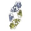
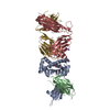
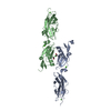
protein localization involved in establishment of planar polarity / apical constriction / establishment of planar polarity of embryonic epithelium / establishment or maintenance of actin cytoskeleton polarity / embryonic morphogenesis / melanosome organization / protein localization to adherens junction / actomyosin structure organization / cortical actin cytoskeleton / apical junction complex ...protein localization involved in establishment of planar polarity / apical constriction / establishment of planar polarity of embryonic epithelium / establishment or maintenance of actin cytoskeleton polarity / embryonic morphogenesis / melanosome organization / protein localization to adherens junction / actomyosin structure organization / cortical actin cytoskeleton / apical junction complex / bicellular tight junction / actin filament organization / adherens junction / cell morphogenesis / kinase binding / cell-cell junction / actin filament binding / cell migration / actin binding / actin cytoskeleton organization / cytoskeleton / microtubule / apical plasma membrane / protein homodimerization activity / plasma membrane Similarity search - Function
Single alpha-helices involved in coiled-coils or other helix-helix interfaces - #3120 / Apx/Shrm Domain 2 / Shroom family / Apx/Shroom domain ASD2 / ASD2 domain profile. / Single alpha-helices involved in coiled-coils or other helix-helix interfaces / Helix non-globular / Special Similarity search - Domain/homology
Redundancy: 10 % / Av σ(I) over netI: 36.18 / Number: 76057 / Rmerge(I) obs: 0.085 / Χ2: 3.25 / D res high: 3.5 Å / D res low: 30 Å / Num. obs: 7573 / % possible obs: 99.3
In the structure databanks used in Yorodumi, some data are registered as the other names, "COVID-19 virus" and "2019-nCoV". Here are the details of the virus and the list of structure data.
Jan 31, 2019. EMDB accession codes are about to change! (news from PDBe EMDB page)
EMDB accession codes are about to change! (news from PDBe EMDB page)
The allocation of 4 digits for EMDB accession codes will soon come to an end. Whilst these codes will remain in use, new EMDB accession codes will include an additional digit and will expand incrementally as the available range of codes is exhausted. The current 4-digit format prefixed with “EMD-” (i.e. EMD-XXXX) will advance to a 5-digit format (i.e. EMD-XXXXX), and so on. It is currently estimated that the 4-digit codes will be depleted around Spring 2019, at which point the 5-digit format will come into force.
The EM Navigator/Yorodumi systems omit the EMD- prefix.
Related info.:Q: What is EMD? / ID/Accession-code notation in Yorodumi/EM Navigator
Yorodumi is a browser for structure data from EMDB, PDB, SASBDB, etc.
This page is also the successor to EM Navigator detail page, and also detail information page/front-end page for Omokage search.
The word "yorodu" (or yorozu) is an old Japanese word meaning "ten thousand". "mi" (miru) is to see.
Related info.:EMDB / PDB / SASBDB / Comparison of 3 databanks / Yorodumi Search / Aug 31, 2016. New EM Navigator & Yorodumi / Yorodumi Papers / Jmol/JSmol / Function and homology information / Changes in new EM Navigator and Yorodumi
 Movie
Movie Controller


 Open data
 Basic information
 Components
 Keywords
 Function and homology information
 X-RAY DIFFRACTION /
  Authors
 Citation
 Structure visualization
 Downloads & links
 Other downloads
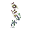


 PDBj
PDBj Assembly

 Sample preparation
 Processing