 Movie
Movie Controller

[English] 日本語
 Yorodumi
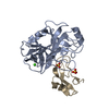
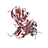
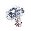
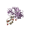
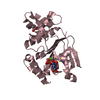
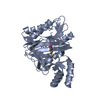
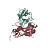
- PDB-3tgk: TRYPSINOGEN MUTANT D194N AND DELETION OF ILE 16-VAL 17 COMPLEXED ... -
+ Open data

- Basic information
| Entry | Database: PDB / ID: 3tgk | ||||||
|---|---|---|---|---|---|---|---|
| Title | TRYPSINOGEN MUTANT D194N AND DELETION OF ILE 16-VAL 17 COMPLEXED WITH BOVINE PANCREATIC TRYPSIN INHIBITOR (BPTI) | ||||||
 Components Components |
| ||||||
 Keywords Keywords | HYDROLASE/ HYDROLASE INHIBITOR / SERINE PROTEASE / COMPLEX (SERINE PROTEASE-INHIBITOR) / HYDROLASE- HYDROLASE INHIBITOR COMPLEX | ||||||
| Function / homology |  Function and homology information Function and homology informationAntimicrobial peptides / Alpha-defensins / Activation of Matrix Metalloproteinases / trypsinogen activation / negative regulation of serine-type endopeptidase activity / sulfate binding / potassium channel inhibitor activity / negative regulation of platelet aggregation / zymogen binding / Neutrophil degranulation ...Antimicrobial peptides / Alpha-defensins / Activation of Matrix Metalloproteinases / trypsinogen activation / negative regulation of serine-type endopeptidase activity / sulfate binding / potassium channel inhibitor activity / negative regulation of platelet aggregation / zymogen binding / Neutrophil degranulation / molecular function inhibitor activity / negative regulation of thrombin-activated receptor signaling pathway / collagen catabolic process / trypsin / serine protease inhibitor complex / digestion / response to nutrient / serine-type endopeptidase inhibitor activity / protease binding / serine-type endopeptidase activity / calcium ion binding / proteolysis / extracellular space / extracellular region Similarity search - Function | ||||||
| Biological species |  | ||||||
| Method |  X-RAY DIFFRACTION / OTHER / Resolution: 1.7 Å X-RAY DIFFRACTION / OTHER / Resolution: 1.7 Å | ||||||
 Authors Authors | Pasternak, A. / White, A. / Jeffery, C.J. / Medina, N. / Cahoon, M. / Ringe, D. / Hedstrom, L. | ||||||
 Citation Citation | Journal: Protein Sci. / Year: 2001 Title: The energetic cost of induced fit catalysis: Crystal structures of trypsinogen mutants with enhanced activity and inhibitor affinity. Authors: Pasternak, A. / White, A. / Jeffery, C.J. / Medina, N. / Cahoon, M. / Ringe, D. / Hedstrom, L. | ||||||
| History |
|
- Structure visualization
| Structure viewer | Molecule: MolmilJmol/JSmol |
|---|
- Downloads & links
-Download
| PDBx/mmCIF format | 3tgk.cif.gz | 72.8 KB | Display | PDBx/mmCIF format |
|---|---|---|---|---|
| PDB format | pdb3tgk.ent.gz | 52 KB | Display | PDB format |
| PDBx/mmJSON format | 3tgk.json.gz | Tree view | PDBx/mmJSON format | |
| Others |  Other downloads Other downloads |
-Validation report
| Summary document | 3tgk_validation.pdf.gz | 443.5 KB | Display | wwPDB validaton report |
|---|---|---|---|---|
| Full document | 3tgk_full_validation.pdf.gz | 443.9 KB | Display | |
| Data in XML | 3tgk_validation.xml.gz | 14.4 KB | Display | |
| Data in CIF | 3tgk_validation.cif.gz | 20.3 KB | Display | |
| Arichive directory | https://data.pdbj.org/pub/pdb/validation_reports/tg/3tgkftp://data.pdbj.org/pub/pdb/validation_reports/tg/3tgk | HTTPS FTP |
-Related structure data


-Links
 PDBj
PDBj


- Assembly
| Deposited unit | 
| ||||||||
|---|---|---|---|---|---|---|---|---|---|
| 1 |
| ||||||||
| Unit cell |
|
-Components
| #1: Protein | Mass: 24734.705 Da / Num. of mol.: 1 / Mutation: DEL(I16,V17), D194N Source method: isolated from a genetically manipulated source Source: (gene. exp.)  | ||||
|---|---|---|---|---|---|
| #2: Protein | Mass: 7337.477 Da / Num. of mol.: 1 / Source method: isolated from a natural source / Source: (natural) | ||||
| #3: Chemical | ChemComp-CA /   Mass: 40.078 Da / Num. of mol.: 1 / Source method: obtained synthetically / Formula: Ca Mass: 40.078 Da / Num. of mol.: 1 / Source method: obtained synthetically / Formula: Ca | ||||
| #4: Chemical |   Mass: 96.063 Da / Num. of mol.: 3 / Source method: obtained synthetically / Formula: SO4 Mass: 96.063 Da / Num. of mol.: 3 / Source method: obtained synthetically / Formula: SO4#5: Water | ChemComp-HOH / |  Mass: 18.015 Da / Num. of mol.: 210 / Source method: isolated from a natural source / Formula: H2O Mass: 18.015 Da / Num. of mol.: 210 / Source method: isolated from a natural source / Formula: H2OHas protein modification | Y | |
-Experimental details
-Experiment
| Experiment | Method: X-RAY DIFFRACTION / Number of used crystals: 1 |
|---|
- Sample preparation
| Crystal | Density Matthews: 2.39 Å3/Da / Density % sol: 48.56 % | ||||||||||||||||||||||||||||||||||||||||||||||||
|---|---|---|---|---|---|---|---|---|---|---|---|---|---|---|---|---|---|---|---|---|---|---|---|---|---|---|---|---|---|---|---|---|---|---|---|---|---|---|---|---|---|---|---|---|---|---|---|---|---|
| Crystal grow | pH: 8.5 / Details: 0.2 M LISO4, 0.1 M TRIS, pH 8.5 | ||||||||||||||||||||||||||||||||||||||||||||||||
| Crystal grow | *PLUS Method: vapor diffusion, hanging drop | ||||||||||||||||||||||||||||||||||||||||||||||||
| Components of the solutions | *PLUS
|
-Data collection
| Diffraction | Mean temperature: 277 K |
|---|---|
| Diffraction source | Source: ROTATING ANODE / Type: RIGAKU RU200 / Wavelength: 1.5418 |
| Detector | Type: RIGAKU RAXIS IIC / Detector: IMAGE PLATE |
| Radiation | Protocol: SINGLE WAVELENGTH / Monochromatic (M) / Laue (L): M / Scattering type: x-ray |
| Radiation wavelength | Wavelength: 1.5418 Å / Relative weight: 1 |
| Reflection | Resolution: 1.7→20 Å / Num. obs: 32654 / % possible obs: 95.8 % / Redundancy: 5.5 % / Rsym value: 0.057 / Net I/σ(I): 10 |
| Reflection shell | Resolution: 1.7→1.76 Å / % possible all: 94.2 |
| Reflection | *PLUS Num. measured all: 181142 / Rmerge(I) obs: 0.057 |
- Processing
| Software |
| ||||||||||||||||||||||||||||||||||||||||||||||||||||||||||||
|---|---|---|---|---|---|---|---|---|---|---|---|---|---|---|---|---|---|---|---|---|---|---|---|---|---|---|---|---|---|---|---|---|---|---|---|---|---|---|---|---|---|---|---|---|---|---|---|---|---|---|---|---|---|---|---|---|---|---|---|---|---|
| Refinement | Method to determine structure: OTHER / Resolution: 1.7→10 Å / Isotropic thermal model: RESTRAINED / Cross valid method: THROUGHOUT / σ(F): 0 / Details: ALSO TOPH19.PEP USED
| ||||||||||||||||||||||||||||||||||||||||||||||||||||||||||||
| Displacement parameters | Biso mean: 17.3 Å2 | ||||||||||||||||||||||||||||||||||||||||||||||||||||||||||||
| Refinement step | Cycle: LAST / Resolution: 1.7→10 Å
| ||||||||||||||||||||||||||||||||||||||||||||||||||||||||||||
| Refine LS restraints |
| ||||||||||||||||||||||||||||||||||||||||||||||||||||||||||||
| LS refinement shell | Resolution: 1.7→1.78 Å / Total num. of bins used: 8 /
| ||||||||||||||||||||||||||||||||||||||||||||||||||||||||||||
| Xplor file |
| ||||||||||||||||||||||||||||||||||||||||||||||||||||||||||||
| Software | *PLUS Name: X-PLOR / Version: 3.851 / Classification: refinement | ||||||||||||||||||||||||||||||||||||||||||||||||||||||||||||
| Refinement | *PLUS Highest resolution: 1.7 Å / Lowest resolution: 10 Å / σ(F): 0 / % reflection Rfree: 10 % | ||||||||||||||||||||||||||||||||||||||||||||||||||||||||||||
| Solvent computation | *PLUS | ||||||||||||||||||||||||||||||||||||||||||||||||||||||||||||
| Displacement parameters | *PLUS Biso mean: 17.3 Å2 | ||||||||||||||||||||||||||||||||||||||||||||||||||||||||||||
| Refine LS restraints | *PLUS
| ||||||||||||||||||||||||||||||||||||||||||||||||||||||||||||
| LS refinement shell | *PLUS Rfactor Rfree: 0.264 / Rfactor Rwork: 0.262 |
