 Movie
Movie Controller

[English] 日本語
 Yorodumi
- PDB-3tfy: Naa50p amino-terminal acetyltransferase bound to substrate peptid... -
+ Open data

- Basic information
| Entry | Database: PDB / ID: 3tfy | ||||||
|---|---|---|---|---|---|---|---|
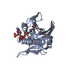
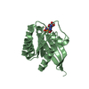
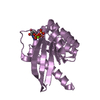
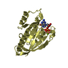
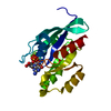
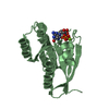
| Title | Naa50p amino-terminal acetyltransferase bound to substrate peptide fragment and CoA | ||||||
 Components Components |
| ||||||
 Keywords Keywords | TRANSFERASE / GNAT Family / N(alpha)-acetyltransferase | ||||||
| Function / homology |  Function and homology information Function and homology informationmitotic sister chromatid cohesion, centromeric / N-terminal methionine Nalpha-acetyltransferase NatE / N-terminal protein amino acid acetylation / NatA complex / protein N-terminal-methionine acetyltransferase activity / protein-N-terminal amino-acid acetyltransferase activity / histone H4 acetyltransferase activity / establishment of mitotic sister chromatid cohesion / FGFR2 alternative splicing / mitotic sister chromatid cohesion ...mitotic sister chromatid cohesion, centromeric / N-terminal methionine Nalpha-acetyltransferase NatE / N-terminal protein amino acid acetylation / NatA complex / protein N-terminal-methionine acetyltransferase activity / protein-N-terminal amino-acid acetyltransferase activity / histone H4 acetyltransferase activity / establishment of mitotic sister chromatid cohesion / FGFR2 alternative splicing / mitotic sister chromatid cohesion / regulation of RNA splicing / Processing of Capped Intron-Containing Pre-mRNA / protein-lysine-acetyltransferase activity / RNA processing / catalytic step 2 spliceosome / mRNA Splicing - Major Pathway / Transferases; Acyltransferases; Transferring groups other than aminoacyl groups / post-translational protein modification / Gene and protein expression by JAK-STAT signaling after Interleukin-12 stimulation / mRNA splicing, via spliceosome / single-stranded RNA binding / ribonucleoprotein complex / synapse / nucleolus / RNA binding / extracellular exosome / nucleoplasm / membrane / nucleus / plasma membrane / cytoplasm / cytosol Similarity search - Function | ||||||
| Biological species |  Homo sapiens (human) Homo sapiens (human) | ||||||
| Method |  X-RAY DIFFRACTION / SYNCHROTRON / MOLECULAR REPLACEMENT / Resolution: 2.75 Å X-RAY DIFFRACTION / SYNCHROTRON / MOLECULAR REPLACEMENT / Resolution: 2.75 Å | ||||||
 Authors Authors | Liszczak, G.P. / Marmorstein, R. | ||||||
 Citation Citation | Journal: J.Biol.Chem. / Year: 2011 Title: Structure of a Ternary Naa50p (NAT5/SAN) N-terminal Acetyltransferase Complex Reveals the Molecular Basis for Substrate-specific Acetylation. Authors: Liszczak, G. / Arnesen, T. / Marmorstein, R. | ||||||
| History |
|
- Structure visualization
| Structure viewer | Molecule: MolmilJmol/JSmol |
|---|
- Downloads & links
-Download
| PDBx/mmCIF format | 3tfy.cif.gz | 113.4 KB | Display | PDBx/mmCIF format |
|---|---|---|---|---|
| PDB format | pdb3tfy.ent.gz | 87.5 KB | Display | PDB format |
| PDBx/mmJSON format | 3tfy.json.gz | Tree view | PDBx/mmJSON format | |
| Others |  Other downloads Other downloads |
-Validation report
| Arichive directory | https://data.pdbj.org/pub/pdb/validation_reports/tf/3tfyftp://data.pdbj.org/pub/pdb/validation_reports/tf/3tfy | HTTPS FTP |
|---|
-Related structure data
| Related structure data |  2pswS S: Starting model for refinement |
|---|---|
| Similar structure data |


-Links
 PDBj
PDBj


- Assembly
| Deposited unit | 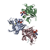
| ||||||||
|---|---|---|---|---|---|---|---|---|---|
| 1 | 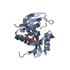
| ||||||||
| 2 | 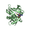
| ||||||||
| 3 | 
| ||||||||
| Unit cell |
|
-Components
| #1: Protein | Mass: 19427.373 Da / Num. of mol.: 3 Source method: isolated from a genetically manipulated source Source: (gene. exp.) Homo sapiens (human) / Gene: MAK3, NAA50, NAT13, NAT5 / Plasmid: pETM-GST / Production host:  References: UniProt: Q9GZZ1, Transferases; Acyltransferases; Transferring groups other than aminoacyl groups #2: Protein/peptide | Mass: 1060.208 Da / Num. of mol.: 3 / Source method: obtained synthetically / References: UniProt: P52597*PLUS #3: Chemical |   Mass: 767.534 Da / Num. of mol.: 3 / Source method: obtained synthetically / Formula: C21H36N7O16P3S Mass: 767.534 Da / Num. of mol.: 3 / Source method: obtained synthetically / Formula: C21H36N7O16P3S#4: Water | ChemComp-HOH / |  Mass: 18.015 Da / Num. of mol.: 93 / Source method: isolated from a natural source / Formula: H2O Mass: 18.015 Da / Num. of mol.: 93 / Source method: isolated from a natural source / Formula: H2O |
|---|
-Experimental details
-Experiment
| Experiment | Method: X-RAY DIFFRACTION / Number of used crystals: 1 |
|---|
- Sample preparation
| Crystal | Density Matthews: 2.68 Å3/Da / Density % sol: 54.09 % |
|---|---|
| Crystal grow | Temperature: 273 K / Method: vapor diffusion, hanging drop / pH: 5 Details: well solution: 16% PEG 8000, 20% glycerol, 40mM potassium phosphate (monobasic), pH 5.0, VAPOR DIFFUSION, HANGING DROP, temperature 273K |
-Data collection
| Diffraction | Mean temperature: 100 K |
|---|---|
| Diffraction source | Source: SYNCHROTRON / Site: NSLS  / Beamline: X6A / Wavelength: 1.1271 Å / Beamline: X6A / Wavelength: 1.1271 Å |
| Detector | Type: ADSC QUANTUM 270 / Detector: CCD / Date: Jun 10, 2010 / Details: Toroidal focusing mirror |
| Radiation | Monochromator: Si(111) channel cut monochromator / Protocol: SINGLE WAVELENGTH / Monochromatic (M) / Laue (L): M / Scattering type: x-ray |
| Radiation wavelength | Wavelength: 1.1271 Å / Relative weight: 1 |
| Reflection | Resolution: 2.75→50 Å / Num. all: 17043 / Num. obs: 16856 / % possible obs: 98.9 % / Observed criterion σ(I): -3 / Redundancy: 3.6 % / Rsym value: 12.9 / Net I/σ(I): 10.2 |
| Reflection shell | Resolution: 2.75→2.8 Å / Redundancy: 3.2 % / Mean I/σ(I) obs: 2.1 / Num. unique all: 682 / Rsym value: 40.9 / % possible all: 93.2 |
- Processing
| Software |
| |||||||||||||||||||||||||||||||||||||||||||||||||
|---|---|---|---|---|---|---|---|---|---|---|---|---|---|---|---|---|---|---|---|---|---|---|---|---|---|---|---|---|---|---|---|---|---|---|---|---|---|---|---|---|---|---|---|---|---|---|---|---|---|---|
| Refinement | Method to determine structure: MOLECULAR REPLACEMENT Starting model: PDB entry 2PSW (cofactor and solvent removed) Resolution: 2.75→33.659 Å / SU ML: 0.39 / σ(F): 0.19 / Phase error: 27.65 / Stereochemistry target values: ML
| |||||||||||||||||||||||||||||||||||||||||||||||||
| Solvent computation | Shrinkage radii: 0.9 Å / VDW probe radii: 1.11 Å / Solvent model: FLAT BULK SOLVENT MODEL / Bsol: 39.837 Å2 / ksol: 0.315 e/Å3 | |||||||||||||||||||||||||||||||||||||||||||||||||
| Displacement parameters |
| |||||||||||||||||||||||||||||||||||||||||||||||||
| Refinement step | Cycle: LAST / Resolution: 2.75→33.659 Å
| |||||||||||||||||||||||||||||||||||||||||||||||||
| Refine LS restraints |
| |||||||||||||||||||||||||||||||||||||||||||||||||
| LS refinement shell |
|
