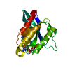
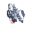
- PDB-1oix: X-ray structure of the small G protein Rab11a in complex with GDP... -
+
Open data
ID or keywords:
Loading...
-
Basic information
Entry
Database: PDB / ID: 1oix
Title
X-ray structure of the small G protein Rab11a in complex with GDP and Pi
Components
RAS-RELATED PROTEIN RAB-11A
Keywords
PROTEIN TRANSPORT / SMALL G PROTEIN / INTRACELLULAR TRAFFICKING / GTP-BINDING / LIPOPROTEIN / PRENYLATION
Function / homology
Function and homology information
: / Anchoring of the basal body to the plasma membrane / VxPx cargo-targeting to cilium / RAB geranylgeranylation / : / regulation of protein localization to centrosome / synaptic vesicle endosomal processing / : / regulation of endocytic recycling / establishment of protein localization to organelle ...: / Anchoring of the basal body to the plasma membrane / VxPx cargo-targeting to cilium / RAB geranylgeranylation / : / regulation of protein localization to centrosome / synaptic vesicle endosomal processing / : / regulation of endocytic recycling / establishment of protein localization to organelle / postsynaptic recycling endosome / positive regulation of mitotic cytokinetic process / establishment of vesicle localization / regulation of protein transport / regulation of cilium assembly / amyloid-beta clearance by transcytosis / vesicle-mediated transport in synapse / neurotransmitter receptor transport, endosome to postsynaptic membrane / exosomal secretion / kinetochore microtubule / presynaptic endosome / VxPx cargo-targeting to cilium / plasma membrane to endosome transport / exocytic vesicle / regulation of vesicle-mediated transport / protein transmembrane transport / astral microtubule organization / RAB geranylgeranylation / myosin V binding / multivesicular body assembly / protein localization to cilium / melanosome transport / Golgi to plasma membrane protein transport / establishment of protein localization to membrane / TBC/RABGAPs / protein localization to cell surface / syntaxin binding / dynein light intermediate chain binding / mitotic metaphase chromosome alignment / exocytosis / cleavage furrow / positive regulation of epithelial cell migration / mitotic spindle assembly / positive regulation of G2/M transition of mitotic cell cycle / positive regulation of axon extension / transport vesicle / phagocytic vesicle / multivesicular body / vesicle-mediated transport / Anchoring of the basal body to the plasma membrane / cytoplasmic vesicle membrane / trans-Golgi network membrane / regulation of cytokinesis / small monomeric GTPase / protein localization to plasma membrane / positive regulation of protein localization to plasma membrane / Translocation of SLC2A4 (GLUT4) to the plasma membrane / trans-Golgi network / centriole / recycling endosome / regulation of long-term neuronal synaptic plasticity / Schaffer collateral - CA1 synapse / recycling endosome membrane / centriolar satellite / spindle pole / neuron projection development / endocytic vesicle membrane / Vasopressin regulates renal water homeostasis via Aquaporins / synaptic vesicle membrane / G protein activity / cytoplasmic vesicle / microtubule binding / vesicle / endosome / Golgi membrane / protein domain specific binding / axon / GTPase activity / centrosome / GTP binding / perinuclear region of cytoplasm / glutamatergic synapse / Golgi apparatus / protein-containing complex / extracellular exosome / cytosol Similarity search - Function
: / Small GTPase Rab domain profile. / Ran (Ras-related nuclear proteins) /TC4 subfamily of small GTPases / Rho (Ras homology) subfamily of Ras-like small GTPases / Ras subfamily of RAS small GTPases / Small GTPase / Ras family / Rab subfamily of small GTPases / Small GTP-binding protein domain / P-loop containing nucleotide triphosphate hydrolases ...: / Small GTPase Rab domain profile. / Ran (Ras-related nuclear proteins) /TC4 subfamily of small GTPases / Rho (Ras homology) subfamily of Ras-like small GTPases / Ras subfamily of RAS small GTPases / Small GTPase / Ras family / Rab subfamily of small GTPases / Small GTP-binding protein domain / P-loop containing nucleotide triphosphate hydrolases / Rossmann fold / P-loop containing nucleoside triphosphate hydrolase / 3-Layer(aba) Sandwich / Alpha Beta Similarity search - Domain/homology
GUANOSINE-5'-DIPHOSPHATE / PHOSPHATE ION / Ras-related protein Rab-11A / Ras-related protein Rab-11A Similarity search - Component
Mass: 18.015 Da / Num. of mol.: 163 / Source method: isolated from a natural source / Formula: H2O
-
Details
Compound details
ENGINEERED MUTATION GLN 70 LEU IN CHAIN A
Sequence details
RESIDUES OF THE N-TERMINAL HIS6-TAG AND LINKER WERE DISORDERED AND NOT VISIBLE IN THE ELECTRON ...RESIDUES OF THE N-TERMINAL HIS6-TAG AND LINKER WERE DISORDERED AND NOT VISIBLE IN THE ELECTRON DENSITY MAP, AS WELL AS THE FIRST 5 RESIDUES OF RAB11A. ASP6 AND GLU7 WERE MODELLED AS ALANINES BECAUSE SIDE CHAINS WERE NOT VISIBLE.
-
Experimental details
-
Experiment
Experiment
Method: X-RAY DIFFRACTION / Number of used crystals: 1
-
Sample preparation
Crystal
Density Matthews: 1.99 Å3/Da / Density % sol: 38.19 %
Crystal grow
pH: 6.5 Details: 1.4 M NACL, 0.15 M NAH2PO4, 0.15 M KH2PO4,0.1 M NAMES PH6.5, pH 6.50
Resolution: 1.7→30 Å / Rfactor Rfree error: 0.006 / Cross valid method: THROUGHOUT / σ(F): 0 Details: FINAL ROUND OF REFINEMENT WAS CARRIED OUT WITH REFMAC
In the structure databanks used in Yorodumi, some data are registered as the other names, "COVID-19 virus" and "2019-nCoV". Here are the details of the virus and the list of structure data.
Jan 31, 2019. EMDB accession codes are about to change! (news from PDBe EMDB page)
EMDB accession codes are about to change! (news from PDBe EMDB page)
The allocation of 4 digits for EMDB accession codes will soon come to an end. Whilst these codes will remain in use, new EMDB accession codes will include an additional digit and will expand incrementally as the available range of codes is exhausted. The current 4-digit format prefixed with “EMD-” (i.e. EMD-XXXX) will advance to a 5-digit format (i.e. EMD-XXXXX), and so on. It is currently estimated that the 4-digit codes will be depleted around Spring 2019, at which point the 5-digit format will come into force.
The EM Navigator/Yorodumi systems omit the EMD- prefix.
Related info.:Q: What is EMD? / ID/Accession-code notation in Yorodumi/EM Navigator
Yorodumi is a browser for structure data from EMDB, PDB, SASBDB, etc.
This page is also the successor to EM Navigator detail page, and also detail information page/front-end page for Omokage search.
The word "yorodu" (or yorozu) is an old Japanese word meaning "ten thousand". "mi" (miru) is to see.
Related info.:EMDB / PDB / SASBDB / Comparison of 3 databanks / Yorodumi Search / Aug 31, 2016. New EM Navigator & Yorodumi / Yorodumi Papers / Jmol/JSmol / Function and homology information / Changes in new EM Navigator and Yorodumi
 Movie
Movie Controller

 Yorodumi
 Open data
 Basic information
 Components
 Keywords
 Function and homology information
 HOMO SAPIENS (human)
 X-RAY DIFFRACTION /
  Authors
 Citation
 Structure visualization
 Downloads & links
 Other downloads
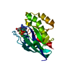


 PDBj
PDBj


 Assembly


 Type: RNA linking / Mass: 443.201 Da / Num. of mol.: 1 / Source method: obtained synthetically / Formula: C10H15N5O11P2 / Comment: GDP, energy-carrying molecule*YM
 Mass: 94.971 Da / Num. of mol.: 1 / Source method: obtained synthetically / Formula: PO4
 Mass: 24.305 Da / Num. of mol.: 1 / Source method: obtained synthetically / Formula: Mg
 Mass: 35.453 Da / Num. of mol.: 1 / Source method: obtained synthetically / Formula: Cl
 Sample preparation
 / Beamline: ID14-1 / Wavelength: 0.934
  Processing