 Movie
Movie Controller

[English] 日本語
 Yorodumi
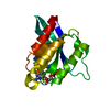
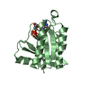
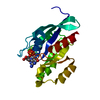
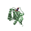
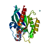
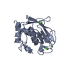
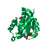
- PDB-1oiw: X-ray structure of the small G protein Rab11a in complex with GTP... -
+ Open data

- Basic information
| Entry | Database: PDB / ID: 1oiw | ||||||
|---|---|---|---|---|---|---|---|
| Title | X-ray structure of the small G protein Rab11a in complex with GTPgammaS | ||||||
 Components Components | RAS-RELATED PROTEIN RAB-11A | ||||||
 Keywords Keywords | PROTEIN TRANSPORT / SMALL G PROTEIN / INTRACELLULAR TRAFFICKING / GTP-BINDING / LIPOPROTEIN / PRENYLATION | ||||||
| Function / homology |  Function and homology information Function and homology information: / Anchoring of the basal body to the plasma membrane / VxPx cargo-targeting to cilium / RAB geranylgeranylation / : / regulation of protein localization to centrosome / synaptic vesicle endosomal processing / : / regulation of endocytic recycling / vesicle-mediated transport in synapse ...: / Anchoring of the basal body to the plasma membrane / VxPx cargo-targeting to cilium / RAB geranylgeranylation / : / regulation of protein localization to centrosome / synaptic vesicle endosomal processing / : / regulation of endocytic recycling / vesicle-mediated transport in synapse / establishment of protein localization to organelle / postsynaptic recycling endosome / positive regulation of mitotic cytokinetic process / establishment of vesicle localization / regulation of protein transport / regulation of cilium assembly / amyloid-beta clearance by transcytosis / neurotransmitter receptor transport, endosome to postsynaptic membrane / exosomal secretion / kinetochore microtubule / presynaptic endosome / VxPx cargo-targeting to cilium / plasma membrane to endosome transport / exocytic vesicle / regulation of vesicle-mediated transport / protein transmembrane transport / astral microtubule organization / protein localization to cilium / RAB geranylgeranylation / myosin V binding / multivesicular body assembly / melanosome transport / Golgi to plasma membrane protein transport / establishment of protein localization to membrane / TBC/RABGAPs / protein localization to cell surface / syntaxin binding / dynein light intermediate chain binding / exocytosis / mitotic metaphase chromosome alignment / cleavage furrow / positive regulation of epithelial cell migration / mitotic spindle assembly / positive regulation of G2/M transition of mitotic cell cycle / positive regulation of axon extension / transport vesicle / centriolar satellite / phagocytic vesicle / multivesicular body / vesicle-mediated transport / centriole / Anchoring of the basal body to the plasma membrane / cytoplasmic vesicle membrane / protein localization to plasma membrane / trans-Golgi network membrane / regulation of cytokinesis / small monomeric GTPase / positive regulation of protein localization to plasma membrane / Translocation of SLC2A4 (GLUT4) to the plasma membrane / regulation of long-term neuronal synaptic plasticity / trans-Golgi network / recycling endosome / Schaffer collateral - CA1 synapse / recycling endosome membrane / neuron projection development / spindle pole / endocytic vesicle membrane / Vasopressin regulates renal water homeostasis via Aquaporins / synaptic vesicle membrane / G protein activity / cytoplasmic vesicle / microtubule binding / vesicle / endosome / Golgi membrane / protein domain specific binding / axon / GTPase activity / centrosome / GTP binding / perinuclear region of cytoplasm / glutamatergic synapse / Golgi apparatus / protein-containing complex / extracellular exosome / cytosol Similarity search - Function | ||||||
| Biological species |  HOMO SAPIENS (human) HOMO SAPIENS (human) | ||||||
| Method |  X-RAY DIFFRACTION / SYNCHROTRON / MOLECULAR REPLACEMENT / Resolution: 2.05 Å X-RAY DIFFRACTION / SYNCHROTRON / MOLECULAR REPLACEMENT / Resolution: 2.05 Å | ||||||
 Authors Authors | Pasqualato, S. / Senic-Matuglia, F. / Renault, L. / Goud, B. / Salamero, J. / Cherfils, J. | ||||||
 Citation Citation | Journal: J.Biol.Chem. / Year: 2004 Title: The Structural Gdp/GTP Cycle of Rab11 Reveals a Novel Interface Involved in the Dynamics of Recycling Endosomes Authors: Pasqualato, S. / Senic-Matuglia, F. / Renault, L. / Goud, B. / Salamero, J. / Cherfils, J. | ||||||
| History |
|
- Structure visualization
| Structure viewer | Molecule: MolmilJmol/JSmol |
|---|
- Downloads & links
-Download
| PDBx/mmCIF format | 1oiw.cif.gz | 51.4 KB | Display | PDBx/mmCIF format |
|---|---|---|---|---|
| PDB format | pdb1oiw.ent.gz | 35 KB | Display | PDB format |
| PDBx/mmJSON format | 1oiw.json.gz | Tree view | PDBx/mmJSON format | |
| Others |  Other downloads Other downloads |
-Validation report
| Arichive directory | https://data.pdbj.org/pub/pdb/validation_reports/oi/1oiwftp://data.pdbj.org/pub/pdb/validation_reports/oi/1oiw | HTTPS FTP |
|---|
-Related structure data
| Related structure data | 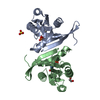 1oivSC S: Starting model for refinement C: citing same article ( |
|---|---|
| Similar structure data |


-Links
 PDBj
PDBj


- Assembly
| Deposited unit | 
| ||||||||||||||||||
|---|---|---|---|---|---|---|---|---|---|---|---|---|---|---|---|---|---|---|---|
| 1 |
| ||||||||||||||||||
| Unit cell |
| ||||||||||||||||||
| Components on special symmetry positions |
|
-Components
| #1: Protein | Mass: 21525.260 Da / Num. of mol.: 1 / Fragment: RESIDUES 1-173 / Mutation: YES Source method: isolated from a genetically manipulated source Details: DELETION MUTANT LACKING THE 43 C-TERMINAL RESIDUES / Source: (gene. exp.) HOMO SAPIENS (human) / Production host:  |
|---|---|
| #2: Chemical | ChemComp-GSP /   Mass: 539.246 Da / Num. of mol.: 1 / Source method: obtained synthetically / Formula: C10H16N5O13P3S Mass: 539.246 Da / Num. of mol.: 1 / Source method: obtained synthetically / Formula: C10H16N5O13P3S |
| #3: Chemical | ChemComp-MG /   Mass: 24.305 Da / Num. of mol.: 1 / Source method: obtained synthetically / Formula: Mg Mass: 24.305 Da / Num. of mol.: 1 / Source method: obtained synthetically / Formula: Mg |
| #4: Water | ChemComp-HOH /  Mass: 18.015 Da / Num. of mol.: 79 / Source method: isolated from a natural source / Formula: H2O Mass: 18.015 Da / Num. of mol.: 79 / Source method: isolated from a natural source / Formula: H2O |
| Compound details | ENGINEERED |
-Experimental details
-Experiment
| Experiment | Method: X-RAY DIFFRACTION / Number of used crystals: 1 |
|---|
- Sample preparation
| Crystal | Density Matthews: 1.98 Å3/Da / Density % sol: 37.73 % | |||||||||||||||||||||||||||||||||||||||||||||||||
|---|---|---|---|---|---|---|---|---|---|---|---|---|---|---|---|---|---|---|---|---|---|---|---|---|---|---|---|---|---|---|---|---|---|---|---|---|---|---|---|---|---|---|---|---|---|---|---|---|---|---|
| Crystal grow | pH: 6.5 Details: 1.4 M NACL, 0.15 M NAH2PO4, 0.15 M KH2PO4,0.1 M NAMES PH6.5, pH 6.50 | |||||||||||||||||||||||||||||||||||||||||||||||||
| Crystal grow | *PLUS Temperature: 20 ℃ / pH: 6.5 / Method: vapor diffusion | |||||||||||||||||||||||||||||||||||||||||||||||||
| Components of the solutions | *PLUS
|
-Data collection
| Diffraction | Mean temperature: 100 K |
|---|---|
| Diffraction source | Source: SYNCHROTRON / Site: ESRF  / Beamline: ID14-2 / Wavelength: 0.934 / Beamline: ID14-2 / Wavelength: 0.934 |
| Detector | Date: May 17, 2002 |
| Radiation | Protocol: SINGLE WAVELENGTH / Monochromatic (M) / Laue (L): M / Scattering type: x-ray |
| Radiation wavelength | Wavelength: 0.934 Å / Relative weight: 1 |
| Reflection | Resolution: 1.9→30 Å / Num. obs: 13509 / % possible obs: 96.5 % / Observed criterion σ(I): 0 / Redundancy: 14.9 % / Biso Wilson estimate: 19.9 Å2 / Rmerge(I) obs: 0.096 / Net I/σ(I): 22.16 |
| Reflection shell | Resolution: 1.9→1.97 Å / Redundancy: 14 % / Rmerge(I) obs: 0.288 / Mean I/σ(I) obs: 9.35 / % possible all: 81.6 |
| Reflection | *PLUS Highest resolution: 1.9 Å / Lowest resolution: 30 Å / Num. obs: 13984 / Num. measured all: 207365 / Rmerge(I) obs: 0.096 |
| Reflection shell | *PLUS % possible obs: 81.6 % / Rmerge(I) obs: 0.288 / Mean I/σ(I) obs: 9.4 |
- Processing
| Software |
| ||||||||||||||||||||||||||||||||||||||||||||||||||||||||||||
|---|---|---|---|---|---|---|---|---|---|---|---|---|---|---|---|---|---|---|---|---|---|---|---|---|---|---|---|---|---|---|---|---|---|---|---|---|---|---|---|---|---|---|---|---|---|---|---|---|---|---|---|---|---|---|---|---|---|---|---|---|---|
| Refinement | Method to determine structure: MOLECULAR REPLACEMENT Starting model: PDB ENTRY 1OIV Resolution: 2.05→30 Å / Rfactor Rfree error: 0.009 / Isotropic thermal model: RESTRAINED / Cross valid method: THROUGHOUT / σ(F): 0 Details: RESIDUES OF THE N-TERMINAL HIS6-TAG AND LINKER WERE DISORDERED AND NOT VISIBLE IN THE ELECTRON DENSITY MAP, AS WELL AS THE FIRST 7 RESIDUES OF RAB11A
| ||||||||||||||||||||||||||||||||||||||||||||||||||||||||||||
| Solvent computation | Solvent model: FLAT MODEL / Bsol: 53.7339 Å2 / ksol: 0.408489 e/Å3 | ||||||||||||||||||||||||||||||||||||||||||||||||||||||||||||
| Displacement parameters | Biso mean: 29.3 Å2
| ||||||||||||||||||||||||||||||||||||||||||||||||||||||||||||
| Refine analyze |
| ||||||||||||||||||||||||||||||||||||||||||||||||||||||||||||
| Refinement step | Cycle: LAST / Resolution: 2.05→30 Å
| ||||||||||||||||||||||||||||||||||||||||||||||||||||||||||||
| Refine LS restraints |
| ||||||||||||||||||||||||||||||||||||||||||||||||||||||||||||
| LS refinement shell | Resolution: 2.05→2.18 Å / Rfactor Rfree error: 0.037 / Total num. of bins used: 6
| ||||||||||||||||||||||||||||||||||||||||||||||||||||||||||||
| Xplor file |
| ||||||||||||||||||||||||||||||||||||||||||||||||||||||||||||
| Refinement | *PLUS Highest resolution: 2.3 Å / Lowest resolution: 30 Å / Num. reflection obs: 8007 / Num. reflection Rfree: 638 / Rfactor Rfree: 0.233 / Rfactor Rwork: 0.225 | ||||||||||||||||||||||||||||||||||||||||||||||||||||||||||||
| Solvent computation | *PLUS | ||||||||||||||||||||||||||||||||||||||||||||||||||||||||||||
| Displacement parameters | *PLUS | ||||||||||||||||||||||||||||||||||||||||||||||||||||||||||||
| Refine LS restraints | *PLUS
| ||||||||||||||||||||||||||||||||||||||||||||||||||||||||||||
| LS refinement shell | *PLUS Highest resolution: 2.3 Å / Lowest resolution: 2.4 Å / Rfactor Rfree: 0.323 / Rfactor Rwork: 0.248 |
