[English] 日本語
Yorodumi
- PDB-3reg: Crystal structure of EhRho1 bound to a GTP analog and Magnesium -

+
Open data


ID or keywords:

Loading...

-
Basic information

Entry
Database: PDB / ID: 3reg
TitleCrystal structure of EhRho1 bound to a GTP analog and Magnesium
ComponentsRho-like small GTPase
KeywordsSIGNALING PROTEIN / cytoskeleton / nucleotide-binding / GTP-binding / lipoprotein / prenylation
Function / homology
Function and homology information


protein localization to phagocytic vesicle / adhesion of symbiont to host cell / phagocytic cup / small GTPase-mediated signal transduction / positive regulation of phagocytosis / phagocytic vesicle / phagocytosis / small monomeric GTPase / cell projection / cytoskeleton ...protein localization to phagocytic vesicle / adhesion of symbiont to host cell / phagocytic cup / small GTPase-mediated signal transduction / positive regulation of phagocytosis / phagocytic vesicle / phagocytosis / small monomeric GTPase / cell projection / cytoskeleton / GTPase activity / GTP binding / magnesium ion binding / plasma membrane
Similarity search - Function
small GTPase Rab1 family profile. / Small GTPase Rho / small GTPase Rho family profile. / small GTPase Ras family profile. / Rho (Ras homology) subfamily of Ras-like small GTPases / Ras subfamily of RAS small GTPases / Small GTPase / Ras family / Rab subfamily of small GTPases / Small GTP-binding protein domain ...small GTPase Rab1 family profile. / Small GTPase Rho / small GTPase Rho family profile. / small GTPase Ras family profile. / Rho (Ras homology) subfamily of Ras-like small GTPases / Ras subfamily of RAS small GTPases / Small GTPase / Ras family / Rab subfamily of small GTPases / Small GTP-binding protein domain / P-loop containing nucleotide triphosphate hydrolases / P-loop containing nucleoside triphosphate hydrolase / Rossmann fold / 3-Layer(aba) Sandwich / Alpha Beta
Similarity search - Domain/homology
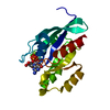
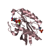
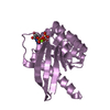
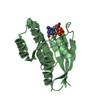
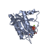
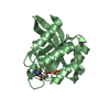
5'-GUANOSINE-DIPHOSPHATE-MONOTHIOPHOSPHATE / Ras-like GTP-binding protein RHO1 / Rho-like small GTPase
Similarity search - Component
Biological speciesEntamoeba histolytica (eukaryote)
MethodX-RAY DIFFRACTION / SYNCHROTRON / MOLECULAR REPLACEMENT / Resolution: 1.801 Å
AuthorsBosch, D.E. / Qiu, C. / Siderovski, D.P.
CitationJournal: J.Biol.Chem. / Year: 2011
Title: Unique structural and nucleotide exchange features of the Rho1 GTPase of Entamoeba histolytica.
Authors: Bosch, D.E. / Wittchen, E.S. / Qiu, C. / Burridge, K. / Siderovski, D.P.
History
DepositionApr 4, 2011Deposition site: RCSB / Processing site: RCSB
Revision 1.0Sep 28, 2011Provider: repository / Type: Initial release
Revision 1.1Mar 14, 2012Group: Database references
Revision 1.2Sep 13, 2023Group: Data collection / Database references ...Data collection / Database references / Derived calculations / Refinement description
Category: chem_comp_atom / chem_comp_bond ...chem_comp_atom / chem_comp_bond / database_2 / pdbx_initial_refinement_model / pdbx_struct_conn_angle / struct_conn / struct_ref_seq_dif / struct_site
Item: _database_2.pdbx_DOI / _database_2.pdbx_database_accession ..._database_2.pdbx_DOI / _database_2.pdbx_database_accession / _pdbx_struct_conn_angle.ptnr1_auth_comp_id / _pdbx_struct_conn_angle.ptnr1_auth_seq_id / _pdbx_struct_conn_angle.ptnr1_label_asym_id / _pdbx_struct_conn_angle.ptnr1_label_atom_id / _pdbx_struct_conn_angle.ptnr1_label_comp_id / _pdbx_struct_conn_angle.ptnr1_label_seq_id / _pdbx_struct_conn_angle.ptnr3_auth_comp_id / _pdbx_struct_conn_angle.ptnr3_auth_seq_id / _pdbx_struct_conn_angle.ptnr3_label_asym_id / _pdbx_struct_conn_angle.ptnr3_label_atom_id / _pdbx_struct_conn_angle.ptnr3_label_comp_id / _pdbx_struct_conn_angle.ptnr3_label_seq_id / _pdbx_struct_conn_angle.value / _struct_conn.pdbx_dist_value / _struct_conn.ptnr1_auth_asym_id / _struct_conn.ptnr1_auth_comp_id / _struct_conn.ptnr1_auth_seq_id / _struct_conn.ptnr1_label_asym_id / _struct_conn.ptnr1_label_atom_id / _struct_conn.ptnr1_label_comp_id / _struct_conn.ptnr1_label_seq_id / _struct_conn.ptnr2_auth_asym_id / _struct_conn.ptnr2_auth_comp_id / _struct_conn.ptnr2_auth_seq_id / _struct_conn.ptnr2_label_asym_id / _struct_conn.ptnr2_label_atom_id / _struct_conn.ptnr2_label_comp_id / _struct_ref_seq_dif.details / _struct_site.pdbx_auth_asym_id / _struct_site.pdbx_auth_comp_id / _struct_site.pdbx_auth_seq_id

-
Structure visualization

Structure viewerMolecule:
MolmilJmol/JSmol

Downloads & links

-
Assembly

Deposited unit
A: Rho-like small GTPase
B: Rho-like small GTPase
hetero molecules


Theoretical massNumber of molelcules
Total (without water)43,9926
Polymers42,8652
Non-polymers1,1274
Water4,125229
1
A: Rho-like small GTPase
hetero molecules


Theoretical massNumber of molelcules
Total (without water)21,9963
Polymers21,4321
Non-polymers5642
Water181
TypeNameSymmetry operationNumber
identity operation1_555x,y,z1
MethodPISA
2
B: Rho-like small GTPase
hetero molecules


Theoretical massNumber of molelcules
Total (without water)21,9963
Polymers21,4321
Non-polymers5642
Water181
TypeNameSymmetry operationNumber
identity operation1_555x,y,z1
MethodPISA
Unit cell
Length a, b, c (Å)36.468, 39.497, 63.578
Angle α, β, γ (deg.)81.75, 80.76, 65.44
Int Tables number1
Space group name H-MP1

-
Components

#1: Protein Rho-like small GTPase


Mass: 21432.455 Da / Num. of mol.: 2 / Fragment: UNP residues 1-191
Source method: isolated from a genetically manipulated source
Source: (gene. exp.) Entamoeba histolytica (eukaryote) / Strain: HM1:IMSS / Gene: EhRho1 / Plasmid: pLIC-His / Production host: Escherichia coli (E. coli) / Strain (production host): BL21 / References: UniProt: Q95TD4, UniProt: C4M4W4*PLUS
#2: Chemical ChemComp-GSP / 5'-GUANOSINE-DIPHOSPHATE-MONOTHIOPHOSPHATE


Mass: 539.246 Da / Num. of mol.: 2 / Source method: obtained synthetically / Formula: C10H16N5O13P3S
#3: Chemical ChemComp-MG / MAGNESIUM ION


Mass: 24.305 Da / Num. of mol.: 2 / Source method: obtained synthetically / Formula: Mg
#4: Water ChemComp-HOH / water / Water


Mass: 18.015 Da / Num. of mol.: 229 / Source method: isolated from a natural source / Formula: H2O

-
Experimental details

-
Experiment

ExperimentMethod: X-RAY DIFFRACTION / Number of used crystals: 1

-
Sample preparation

CrystalDensity Matthews: 1.91 Å3/Da / Density % sol: 35.63 %
Crystal growTemperature: 291 K / Method: vapor diffusion, hanging drop / pH: 4.6
Details: EhRho1 at 15 mg/mL in crystallization buffer (50 mM Tris pH 8.0, 250 mM NaCl, 2.5% (v/v) glycerol, 5 mM DTT, 50 microM GTPgammaS, 1 mM magnesium chloride) was mixed 1:1 with and equilibrated ...Details: EhRho1 at 15 mg/mL in crystallization buffer (50 mM Tris pH 8.0, 250 mM NaCl, 2.5% (v/v) glycerol, 5 mM DTT, 50 microM GTPgammaS, 1 mM magnesium chloride) was mixed 1:1 with and equilibrated against crystallization solution (25% PEG 4000, 150 mM ammonium acetate, 100 mM sodium acetate pH 4.6), VAPOR DIFFUSION, HANGING DROP, temperature 291K

-
Data collection

DiffractionMean temperature: 100 K
Diffraction sourceSource: SYNCHROTRON / Site: APS / Beamline: 22-BM / Wavelength: 1 Å
DetectorType: MAR scanner 300 mm plate / Detector: IMAGE PLATE / Date: Nov 18, 2010 / Details: custom
RadiationMonochromator: Si(111) / Protocol: SINGLE WAVELENGTH / Monochromatic (M) / Laue (L): M / Scattering type: x-ray
Radiation wavelengthWavelength: 1 Å / Relative weight: 1
ReflectionResolution: 1.8→40 Å / Num. all: 29325 / Num. obs: 23929 / % possible obs: 81.6 % / Observed criterion σ(F): 0 / Observed criterion σ(I): 0 / Redundancy: 2 % / Rmerge(I) obs: 0.043 / Net I/σ(I): 16.1
Reflection shellResolution: 1.8→1.82 Å / Redundancy: 1.4 % / Rmerge(I) obs: 0.278 / Mean I/σ(I) obs: 2.8 / % possible all: 45.9

-
Processing

Software
NameVersionClassification
HKL-2000data collection
PHASERphasing
PHENIX(phenix.refine: 1.6_289)refinement
HKL-2000data reduction
HKL-2000data scaling
RefinementMethod to determine structure: MOLECULAR REPLACEMENT
Starting model: PDB ID 1A2B
Resolution: 1.801→32.958 Å / SU ML: 0.22 / σ(F): 1.96 / Phase error: 25.03 / Stereochemistry target values: ML
RfactorNum. reflection% reflection
Rfree0.2221 1214 5.08 %
Rwork0.1718 --
obs0.1744 23882 81.4 %
all-29486 -
Solvent computationShrinkage radii: 0.9 Å / VDW probe radii: 1.11 Å / Solvent model: FLAT BULK SOLVENT MODEL / Bsol: 41.113 Å2 / ksol: 0.352 e/Å3
Displacement parameters
Baniso -1Baniso -2Baniso -3
1-0.3683 Å20.703 Å2-0.5607 Å2
2---1.8001 Å22.6031 Å2
3---1.4318 Å2
Refinement stepCycle: LAST / Resolution: 1.801→32.958 Å
ProteinNucleic acidLigandSolventTotal
Num. atoms2658 0 66 229 2953
Refine LS restraints
Refine-IDTypeDev idealNumber
X-RAY DIFFRACTIONf_bond_d0.0092775
X-RAY DIFFRACTIONf_angle_d1.23770
X-RAY DIFFRACTIONf_dihedral_angle_d13.8951004
X-RAY DIFFRACTIONf_chiral_restr0.078431
X-RAY DIFFRACTIONf_plane_restr0.005465
LS refinement shell
Resolution (Å)Rfactor RfreeNum. reflection RfreeRfactor RworkNum. reflection RworkRefine-ID% reflection obs (%)
1.801-1.87350.2907860.21541558X-RAY DIFFRACTION50
1.8735-1.95880.29941050.20291931X-RAY DIFFRACTION62
1.9588-2.0620.24541260.19852250X-RAY DIFFRACTION73
2.062-2.19120.25121230.18582480X-RAY DIFFRACTION80
2.1912-2.36030.23391240.18142679X-RAY DIFFRACTION87
2.3603-2.59780.23341460.18212863X-RAY DIFFRACTION92
2.5978-2.97350.24131700.18342914X-RAY DIFFRACTION95
2.9735-3.74540.1981680.15792998X-RAY DIFFRACTION96
3.7454-32.96370.19281660.15272995X-RAY DIFFRACTION97
Refinement TLS params.

Method: refined / Refine-ID: X-RAY DIFFRACTION

IDL112)L122)L132)L222)L232)L332)S11 (Å °)S12 (Å °)S13 (Å °)S21 (Å °)S22 (Å °)S23 (Å °)S31 (Å °)S32 (Å °)S33 (Å °)T112)T122)T132)T222)T232)T332)Origin x (Å)Origin y (Å)Origin z (Å)
14.9367-0.59223.53374.4703-1.39924.03110.74370.0655-0.225-0.2245-0.07650.4631-0.0229-0.0377-0.56350.6149-0.01570.26280.0741-0.09860.2797-1.8339-24.394547.4485
20.8707-0.0434-0.91190.86410.32622.8793-0.0902-0.02250.18270.04030.06260.2917-0.4391-0.1785-0.00490.19210.0646-0.00510.17380.00420.16275.4396-2.944345.0753
35.3582-4.5507-2.77415.54694.35695.6838-0.2721-0.5693-0.4568-0.02420.13380.6851-0.3516-0.09110.16170.1294-0.0120.05370.263-0.03260.1555-1.1367-9.592147.7797
41.4918-0.7286-0.89390.86950.37020.91760.0092-0.3694-0.09730.0473-0.14050.2227-0.0367-0.14550.11690.16050.02540.03580.24740.01020.10385.1606-9.780351.8831
50.96440.1017-0.06621.05421.20971.55940.0291-0.4448-0.11790.19980.0642-0.23250.15340.4359-0.07510.12660.0138-0.03050.31170.01880.144617.1894-8.71751.7257
62.84050.0088-1.56080.8724-0.12481.6081-0.0164-0.3755-0.1321-0.16060.1253-0.14980.00670.5186-0.08180.1279-0.01380.00120.2366-0.05010.147223.0692-7.661940.1067
71.55381.7258-1.55953.2383-2.76212.858-0.19790.0184-0.3351-0.47450.0289-0.42370.3956-0.13920.14510.13810.00070.00010.1534-0.02060.19659.3375-13.035138.2427
82.6801-2.11020.61792.55731.50724.5357-0.5433-0.105-2.7511-0.05820.9596-0.5478-0.47210.0109-0.15440.4543-0.06020.14430.2935-0.10390.825710.6807-27.596644.4662
94.5466-0.20563.31687.53081.42622.85010.70420.3766-0.520.8886-0.2659-0.01610.74440.4553-0.66940.57120.2849-0.07310.29660.00450.303631.4624-25.711516.857
101.15710.315-0.56210.3865-0.21121.97510.23720.13230.29070.0209-0.1025-0.1721-0.45820.4039-0.11360.1863-0.06770.02940.1272-0.01340.148625.5405-4.836915.7781
112.6311-1.92413.04022.1834-1.92794.62090.38220.1563-0.68710.1167-0.2927-0.16440.37120.7509-0.05450.20910.0575-0.04580.29960.09080.265631.3931-18.903220.2195
121.21640.56930.14630.25730.12630.19650.13710.58320.19460.03790.1238-0.2469-0.27750.0885-0.20010.26160.12050.05030.38140.03920.105222.6003-7.94533.1973
132.0143-0.4102-0.78121.02040.31843.5010.2490.3324-0.1549-0.0641-0.06980.0026-0.1213-0.31550.0550.13310.0474-0.04020.1376-0.03380.098413.3091-10.672812.5713
140.8318-1.8425-1.27435.20441.77574.94550.14520.1467-0.03320.3256-0.0340.5142-0.2448-1.3056-0.12770.19020.1362-0.0040.35950.04490.21674.1762-5.92423.7208
156.31652.75632.83121.57752.28673.01460.36090.0697-0.7470.19470.0292-0.16330.239-0.0956-0.35160.1303-0.0546-0.01920.0760.00840.182113.2568-12.609121.4555
164.1932-2.75760.22792.19510.90276.34210.0538-0.0495-0.77140.13190.11590.91970.79180.3164-0.12980.1874-0.01-0.04990.09880.04860.296521.815-20.662622.1978
Refinement TLS group
IDRefine-IDRefine TLS-IDSelection details
1X-RAY DIFFRACTION1(chain A and resid 2:23)
2X-RAY DIFFRACTION2(chain A and resid 24:56)
3X-RAY DIFFRACTION3(chain A and resid 57:64)
4X-RAY DIFFRACTION4(chain A and resid 65:93)
5X-RAY DIFFRACTION5(chain A and resid 94:135)
6X-RAY DIFFRACTION6(chain A and resid 136:167)
7X-RAY DIFFRACTION7(chain A and resid 168:185)
8X-RAY DIFFRACTION8(chain A and resid 186:191)
9X-RAY DIFFRACTION9(chain B and resid 14:24)
10X-RAY DIFFRACTION10(chain B and resid 25:57)
11X-RAY DIFFRACTION11(chain B and resid 58:75)
12X-RAY DIFFRACTION12(chain B and resid 76:94)
13X-RAY DIFFRACTION13(chain B and resid 95:140)
14X-RAY DIFFRACTION14(chain B and resid 141:154)
15X-RAY DIFFRACTION15(chain B and resid 155:174)
16X-RAY DIFFRACTION16(chain B and resid 175:189)

+
About Yorodumi

-
News

-
Feb 9, 2022. New format data for meta-information of EMDB entries

New format data for meta-information of EMDB entries

  • Version 3 of the EMDB header file is now the official format.
  • The previous official version 1.9 will be removed from the archive.

Related info.:EMDB header

External links:wwPDB to switch to version 3 of the EMDB data model

-
Aug 12, 2020. Covid-19 info

Covid-19 info

URL: https://pdbj.org/emnavi/covid19.php

New page: Covid-19 featured information page in EM Navigator.

Related info.:Covid-19 info / Mar 5, 2020. Novel coronavirus structure data

+
Mar 5, 2020. Novel coronavirus structure data

Novel coronavirus structure data

Related info.:Yorodumi Speices / Aug 12, 2020. Covid-19 info

External links:COVID-19 featured content - PDBj / Molecule of the Month (242):Coronavirus Proteases

+
Jan 31, 2019. EMDB accession codes are about to change! (news from PDBe EMDB page)

EMDB accession codes are about to change! (news from PDBe EMDB page)

  • The allocation of 4 digits for EMDB accession codes will soon come to an end. Whilst these codes will remain in use, new EMDB accession codes will include an additional digit and will expand incrementally as the available range of codes is exhausted. The current 4-digit format prefixed with “EMD-” (i.e. EMD-XXXX) will advance to a 5-digit format (i.e. EMD-XXXXX), and so on. It is currently estimated that the 4-digit codes will be depleted around Spring 2019, at which point the 5-digit format will come into force.
  • The EM Navigator/Yorodumi systems omit the EMD- prefix.

Related info.:Q: What is EMD? / ID/Accession-code notation in Yorodumi/EM Navigator

External links:EMDB Accession Codes are Changing Soon! / Contact to PDBj

+
Jul 12, 2017. Major update of PDB

Major update of PDB

  • wwPDB released updated PDB data conforming to the new PDBx/mmCIF dictionary.
  • This is a major update changing the version number from 4 to 5, and with Remediation, in which all the entries are updated.
  • In this update, many items about electron microscopy experimental information are reorganized (e.g. em_software).
  • Now, EM Navigator and Yorodumi are based on the updated data.

External links:wwPDB Remediation / Enriched Model Files Conforming to OneDep Data Standards Now Available in the PDB FTP Archive

-
Yorodumi

Thousand views of thousand structures

  • Yorodumi is a browser for structure data from EMDB, PDB, SASBDB, etc.
  • This page is also the successor to EM Navigator detail page, and also detail information page/front-end page for Omokage search.
  • The word "yorodu" (or yorozu) is an old Japanese word meaning "ten thousand". "mi" (miru) is to see.

Related info.:EMDB / PDB / SASBDB / Comparison of 3 databanks / Yorodumi Search / Aug 31, 2016. New EM Navigator & Yorodumi / Yorodumi Papers / Jmol/JSmol / Function and homology information / Changes in new EM Navigator and Yorodumi

Read more