 Movie
Movie Controller

[English] 日本語
 Yorodumi
- PDB-3tef: Crystal Structure of the Periplasmic Catecholate-Siderophore Bind... -
+ Open data

- Basic information
| Entry | Database: PDB / ID: 3tef | ||||||
|---|---|---|---|---|---|---|---|
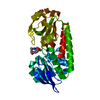
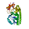
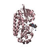
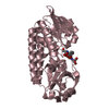
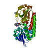
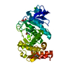
| Title | Crystal Structure of the Periplasmic Catecholate-Siderophore Binding Protein VctP from Vibrio Cholerae | ||||||
 Components Components | Iron(III) ABC transporter, periplasmic iron-compound-binding protein | ||||||
 Keywords Keywords | TRANSPORT PROTEIN / siderophore-binding protein | ||||||
| Function / homology |  Function and homology information Function and homology informationiron coordination entity transport / outer membrane-bounded periplasmic space Similarity search - Function | ||||||
| Biological species |   Vibrio cholerae (bacteria) Vibrio cholerae (bacteria) | ||||||
| Method |  X-RAY DIFFRACTION / SYNCHROTRON / MOLECULAR REPLACEMENT / Resolution: 1.698 Å X-RAY DIFFRACTION / SYNCHROTRON / MOLECULAR REPLACEMENT / Resolution: 1.698 Å | ||||||
 Authors Authors | Liu, X. / Wang, Z. / Liu, S. / Li, N. / Chen, Y. / Zhu, C. / Zhu, D. / Wei, T. / Huang, Y. / Xu, S. / Gu, L. | ||||||
 Citation Citation | Journal: Febs Lett. / Year: 2012 Title: Crystal structure of periplasmic catecholate-siderophore binding protein VctP from Vibrio cholerae at 1.7 A resolution Authors: Liu, X. / Du, Q. / Wang, Z. / Liu, S. / Li, N. / Chen, Y. / Zhu, C. / Zhu, D. / Wei, T. / Huang, Y. / Xu, S. / Gu, L. | ||||||
| History |
|
- Structure visualization
| Structure viewer | Molecule: MolmilJmol/JSmol |
|---|
- Downloads & links
-Download
| PDBx/mmCIF format | 3tef.cif.gz | 71 KB | Display | PDBx/mmCIF format |
|---|---|---|---|---|
| PDB format | pdb3tef.ent.gz | 51.8 KB | Display | PDB format |
| PDBx/mmJSON format | 3tef.json.gz | Tree view | PDBx/mmJSON format | |
| Others |  Other downloads Other downloads |
-Validation report
| Arichive directory | https://data.pdbj.org/pub/pdb/validation_reports/te/3tefftp://data.pdbj.org/pub/pdb/validation_reports/te/3tef | HTTPS FTP |
|---|
-Related structure data
| Similar structure data |
|---|

-Links
 PDBj
PDBj- Assembly
| Deposited unit | 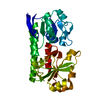
| ||||||||
|---|---|---|---|---|---|---|---|---|---|
| 1 |
| ||||||||
| Unit cell |
|
-Components
| #1: Protein | Mass: 32638.666 Da / Num. of mol.: 1 / Fragment: residues 51-333 Source method: isolated from a genetically manipulated source Source: (gene. exp.) Vibrio cholerae (bacteria) / Gene: VC_A0227 / Production host: |
|---|---|
| #2: Water | ChemComp-HOH /  Mass: 18.015 Da / Num. of mol.: 182 / Source method: isolated from a natural source / Formula: H2O Mass: 18.015 Da / Num. of mol.: 182 / Source method: isolated from a natural source / Formula: H2O |
-Experimental details
-Experiment
| Experiment | Method: X-RAY DIFFRACTION / Number of used crystals: 1 |
|---|
- Sample preparation
| Crystal | Density Matthews: 2.03 Å3/Da / Density % sol: 39.51 % |
|---|---|
| Crystal grow | Temperature: 293 K / Method: vapor diffusion, hanging drop / pH: 8.5 Details: 0.2M ammonium acetate, 0.1M Tris-HCl buffer, 20% PEG3350, pH 8.5, VAPOR DIFFUSION, HANGING DROP, temperature 293K |
-Data collection
| Diffraction | Mean temperature: 100 K |
|---|---|
| Diffraction source | Source: SYNCHROTRON / Site: SSRF  / Beamline: BL17U / Wavelength: 0.9762 Å / Beamline: BL17U / Wavelength: 0.9762 Å |
| Detector | Type: RAYONIX MX-225 / Detector: CCD / Date: Jul 9, 2009 |
| Radiation | Monochromator: Si 111 / Protocol: SINGLE WAVELENGTH / Monochromatic (M) / Laue (L): M / Scattering type: x-ray |
| Radiation wavelength | Wavelength: 0.9762 Å / Relative weight: 1 |
| Reflection | Resolution: 1.698→50 Å / Num. all: 30253 / Num. obs: 30253 / % possible obs: 99.7 % / Observed criterion σ(F): 0 / Observed criterion σ(I): 0 / Redundancy: 8.7 % / Rmerge(I) obs: 0.085 / Rsym value: 0.085 / Net I/σ(I): 23.1 |
| Reflection shell | Resolution: 1.7→1.76 Å / Redundancy: 5.8 % / Rmerge(I) obs: 0.597 / Mean I/σ(I) obs: 2.3 / Num. unique all: 2900 / Rsym value: 0.597 / % possible all: 98.1 |
- Processing
| Software |
| |||||||||||||||||||||||||||||||||||||||||||||||||||||||||||||||||||||||||||||||||||||||||||||||||||||||||
|---|---|---|---|---|---|---|---|---|---|---|---|---|---|---|---|---|---|---|---|---|---|---|---|---|---|---|---|---|---|---|---|---|---|---|---|---|---|---|---|---|---|---|---|---|---|---|---|---|---|---|---|---|---|---|---|---|---|---|---|---|---|---|---|---|---|---|---|---|---|---|---|---|---|---|---|---|---|---|---|---|---|---|---|---|---|---|---|---|---|---|---|---|---|---|---|---|---|---|---|---|---|---|---|---|---|---|
| Refinement | Method to determine structure: MOLECULAR REPLACEMENT / Resolution: 1.698→33.458 Å / Occupancy max: 1 / Occupancy min: 1 / SU ML: 0.17 / σ(F): 0 / Phase error: 22.62 / Stereochemistry target values: ML
| |||||||||||||||||||||||||||||||||||||||||||||||||||||||||||||||||||||||||||||||||||||||||||||||||||||||||
| Solvent computation | Shrinkage radii: 0.83 Å / VDW probe radii: 1.1 Å / Solvent model: FLAT BULK SOLVENT MODEL / Bsol: 40.404 Å2 / ksol: 0.373 e/Å3 | |||||||||||||||||||||||||||||||||||||||||||||||||||||||||||||||||||||||||||||||||||||||||||||||||||||||||
| Displacement parameters | Biso max: 76.61 Å2 / Biso mean: 32.4961 Å2 / Biso min: 10.03 Å2
| |||||||||||||||||||||||||||||||||||||||||||||||||||||||||||||||||||||||||||||||||||||||||||||||||||||||||
| Refinement step | Cycle: LAST / Resolution: 1.698→33.458 Å
| |||||||||||||||||||||||||||||||||||||||||||||||||||||||||||||||||||||||||||||||||||||||||||||||||||||||||
| Refine LS restraints |
| |||||||||||||||||||||||||||||||||||||||||||||||||||||||||||||||||||||||||||||||||||||||||||||||||||||||||
| LS refinement shell | Refine-ID: X-RAY DIFFRACTION / Total num. of bins used: 14
|
