 Movie
Movie Controller


+ Open data

- Basic information
| Entry | Database: PDB / ID: 3t4r | ||||||
|---|---|---|---|---|---|---|---|
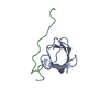
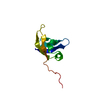
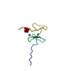
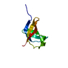
| Title | Lettuce Necrotic Yellow Virus Phosphoprotein C-Terminal Domain | ||||||
 Components Components | Phosphoprotein | ||||||
 Keywords Keywords | VIRAL PROTEIN / Helical bundle / Nucleoprotein | ||||||
| Function / homology | Four Helix Bundle (Hemerythrin (Met), subunit A) - #1590 / : / Lettuce necrotic yellow virus phosphoprotein C-terminal domain / Four Helix Bundle (Hemerythrin (Met), subunit A) / virion component / host cell cytoplasm / Up-down Bundle / Mainly Alpha / Phosphoprotein Function and homology information Function and homology information | ||||||
| Biological species |  Lettuce necrotic yellows virus Lettuce necrotic yellows virus | ||||||
| Method |  X-RAY DIFFRACTION / SYNCHROTRON / SAD / Resolution: 2 Å X-RAY DIFFRACTION / SYNCHROTRON / SAD / Resolution: 2 Å | ||||||
 Authors Authors | Martinez, N. / Tarbouriech, N. / Jamin, M. | ||||||
 Citation Citation | Journal: J.Virol. / Year: 2013 Title: Structure of the C-terminal domain of lettuce necrotic yellows virus phosphoprotein. Authors: Martinez, N. / Ribeiro, E.A. / Leyrat, C. / Tarbouriech, N. / Ruigrok, R.W. / Jamin, M. | ||||||
| History |
|
- Structure visualization
| Structure viewer | Molecule: MolmilJmol/JSmol |
|---|
- Downloads & links
-Download
| PDBx/mmCIF format | 3t4r.cif.gz | 44.6 KB | Display | PDBx/mmCIF format |
|---|---|---|---|---|
| PDB format | pdb3t4r.ent.gz | 32.2 KB | Display | PDB format |
| PDBx/mmJSON format | 3t4r.json.gz | Tree view | PDBx/mmJSON format | |
| Others |  Other downloads Other downloads |
-Validation report
| Arichive directory | https://data.pdbj.org/pub/pdb/validation_reports/t4/3t4rftp://data.pdbj.org/pub/pdb/validation_reports/t4/3t4r | HTTPS FTP |
|---|
-Related structure data
| Similar structure data |
|---|


-Links
 PDBj
PDBj- Assembly
| Deposited unit | 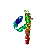
| ||||||||
|---|---|---|---|---|---|---|---|---|---|
| 1 |
| ||||||||
| Unit cell |
| ||||||||
| Components on special symmetry positions |
|
-Components
| #1: Protein | Mass: 9555.391 Da / Num. of mol.: 1 / Fragment: C-terminal domain Source method: isolated from a genetically manipulated source Source: (gene. exp.) Lettuce necrotic yellows virus / Strain: isolate 318 / Gene: P / Plasmid: pET-28a / Production host:  |
|---|---|
| #2: Chemical | ChemComp-MG /   Mass: 24.305 Da / Num. of mol.: 1 / Source method: obtained synthetically / Formula: Mg Mass: 24.305 Da / Num. of mol.: 1 / Source method: obtained synthetically / Formula: Mg |
| #3: Water | ChemComp-HOH /  Mass: 18.015 Da / Num. of mol.: 22 / Source method: isolated from a natural source / Formula: H2O Mass: 18.015 Da / Num. of mol.: 22 / Source method: isolated from a natural source / Formula: H2O |
| Has protein modification | Y |
-Experimental details
-Experiment
| Experiment | Method: X-RAY DIFFRACTION / Number of used crystals: 1 |
|---|
- Sample preparation
| Crystal | Density Matthews: 2.19 Å3/Da / Density % sol: 43.84 % |
|---|---|
| Crystal grow | Temperature: 293 K / Method: vapor diffusion, hanging drop / pH: 5.6 Details: 50 mM Na Cacodylate, 40 mM Magnesium Acetate, 10% MPD, pH 5.6, VAPOR DIFFUSION, HANGING DROP, temperature 293K |
-Data collection
| Diffraction | Mean temperature: 100 K |
|---|---|
| Diffraction source | Source: SYNCHROTRON / Site: ESRF  / Beamline: ID14-4 / Wavelength: 0.974 Å / Beamline: ID14-4 / Wavelength: 0.974 Å |
| Detector | Type: ADSC QUANTUM 315r / Detector: CCD / Date: Apr 10, 2011 |
| Radiation | Monochromator: channel cut ESRF monochromator / Protocol: SINGLE WAVELENGTH / Monochromatic (M) / Laue (L): M / Scattering type: x-ray |
| Radiation wavelength | Wavelength: 0.974 Å / Relative weight: 1 |
| Reflection | Resolution: 2→30.6 Å / Num. obs: 6220 / % possible obs: 99.8 % / Redundancy: 10.56 % / Biso Wilson estimate: 43.8 Å2 / Rmerge(I) obs: 0.072 / Net I/σ(I): 20.62 |
| Reflection shell | Resolution: 2→2.05 Å / Redundancy: 3.7 % / Rmerge(I) obs: 0.455 / Mean I/σ(I) obs: 2.88 / Num. unique all: 813 / % possible all: 99.3 |
- Processing
| Software |
| ||||||||||||||||||||||||||||||||||||||||||||||||||||||||||||||||||||||||||||||||||||||||||||||||||||||||||||||||||||||||||||||||||||||||||||||||||||||
|---|---|---|---|---|---|---|---|---|---|---|---|---|---|---|---|---|---|---|---|---|---|---|---|---|---|---|---|---|---|---|---|---|---|---|---|---|---|---|---|---|---|---|---|---|---|---|---|---|---|---|---|---|---|---|---|---|---|---|---|---|---|---|---|---|---|---|---|---|---|---|---|---|---|---|---|---|---|---|---|---|---|---|---|---|---|---|---|---|---|---|---|---|---|---|---|---|---|---|---|---|---|---|---|---|---|---|---|---|---|---|---|---|---|---|---|---|---|---|---|---|---|---|---|---|---|---|---|---|---|---|---|---|---|---|---|---|---|---|---|---|---|---|---|---|---|---|---|---|---|---|---|
| Refinement | Method to determine structure: SAD / Resolution: 2→30.6 Å / Cor.coef. Fo:Fc: 0.953 / Cor.coef. Fo:Fc free: 0.918 / SU B: 14.241 / SU ML: 0.172 / Cross valid method: THROUGHOUT / ESU R Free: 0.189 / Stereochemistry target values: MAXIMUM LIKELIHOOD / Details: HYDROGENS HAVE BEEN ADDED IN THE RIDING POSITIONS
| ||||||||||||||||||||||||||||||||||||||||||||||||||||||||||||||||||||||||||||||||||||||||||||||||||||||||||||||||||||||||||||||||||||||||||||||||||||||
| Solvent computation | Ion probe radii: 0.8 Å / Shrinkage radii: 0.8 Å / VDW probe radii: 1.4 Å / Solvent model: MASK | ||||||||||||||||||||||||||||||||||||||||||||||||||||||||||||||||||||||||||||||||||||||||||||||||||||||||||||||||||||||||||||||||||||||||||||||||||||||
| Displacement parameters | Biso mean: 54.05 Å2
| ||||||||||||||||||||||||||||||||||||||||||||||||||||||||||||||||||||||||||||||||||||||||||||||||||||||||||||||||||||||||||||||||||||||||||||||||||||||
| Refine analyze |
| ||||||||||||||||||||||||||||||||||||||||||||||||||||||||||||||||||||||||||||||||||||||||||||||||||||||||||||||||||||||||||||||||||||||||||||||||||||||
| Refinement step | Cycle: LAST / Resolution: 2→30.6 Å
| ||||||||||||||||||||||||||||||||||||||||||||||||||||||||||||||||||||||||||||||||||||||||||||||||||||||||||||||||||||||||||||||||||||||||||||||||||||||
| Refine LS restraints |
| ||||||||||||||||||||||||||||||||||||||||||||||||||||||||||||||||||||||||||||||||||||||||||||||||||||||||||||||||||||||||||||||||||||||||||||||||||||||
| LS refinement shell | Resolution: 2→2.05 Å / Total num. of bins used: 20
| ||||||||||||||||||||||||||||||||||||||||||||||||||||||||||||||||||||||||||||||||||||||||||||||||||||||||||||||||||||||||||||||||||||||||||||||||||||||
| Refinement TLS params. | Method: refined / Refine-ID: X-RAY DIFFRACTION
| ||||||||||||||||||||||||||||||||||||||||||||||||||||||||||||||||||||||||||||||||||||||||||||||||||||||||||||||||||||||||||||||||||||||||||||||||||||||
| Refinement TLS group |
|
