- PDB-3t30: Human nucleoplasmin (Npm2): a histone chaperone in oocytes and ea... -
+
Open data
ID or keywords:
Loading...
-
Basic information
Entry
Database: PDB / ID: 3t30
Title
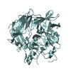
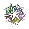
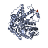
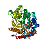
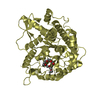
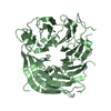
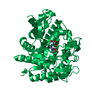
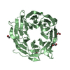
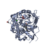
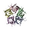
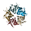
Human nucleoplasmin (Npm2): a histone chaperone in oocytes and early embryos
Components
Nucleoplasmin-2
Keywords
CHAPERONE / beta-barrel jelly roll topology / Histone chaperone / H2A-H2B dimer and H3-H4 tetramer / oocytes and early embryos
Function / homology
Function and homology information
oocyte differentiation / positive regulation of meiotic nuclear division / regulation of exit from mitosis / blastocyst development / single fertilization / Replacement of protamines by nucleosomes in the male pronucleus / positive regulation of DNA replication / histone binding / chromatin remodeling / chromatin binding ...oocyte differentiation / positive regulation of meiotic nuclear division / regulation of exit from mitosis / blastocyst development / single fertilization / Replacement of protamines by nucleosomes in the male pronucleus / positive regulation of DNA replication / histone binding / chromatin remodeling / chromatin binding / chromatin / nucleolus / enzyme binding / RNA binding / nucleoplasm / identical protein binding / nucleus / cytoplasm Similarity search - Function
In the structure databanks used in Yorodumi, some data are registered as the other names, "COVID-19 virus" and "2019-nCoV". Here are the details of the virus and the list of structure data.
Jan 31, 2019. EMDB accession codes are about to change! (news from PDBe EMDB page)
EMDB accession codes are about to change! (news from PDBe EMDB page)
The allocation of 4 digits for EMDB accession codes will soon come to an end. Whilst these codes will remain in use, new EMDB accession codes will include an additional digit and will expand incrementally as the available range of codes is exhausted. The current 4-digit format prefixed with “EMD-” (i.e. EMD-XXXX) will advance to a 5-digit format (i.e. EMD-XXXXX), and so on. It is currently estimated that the 4-digit codes will be depleted around Spring 2019, at which point the 5-digit format will come into force.
The EM Navigator/Yorodumi systems omit the EMD- prefix.
Related info.:Q: What is EMD? / ID/Accession-code notation in Yorodumi/EM Navigator
Yorodumi is a browser for structure data from EMDB, PDB, SASBDB, etc.
This page is also the successor to EM Navigator detail page, and also detail information page/front-end page for Omokage search.
The word "yorodu" (or yorozu) is an old Japanese word meaning "ten thousand". "mi" (miru) is to see.
Related info.:EMDB / PDB / SASBDB / Comparison of 3 databanks / Yorodumi Search / Aug 31, 2016. New EM Navigator & Yorodumi / Yorodumi Papers / Jmol/JSmol / Function and homology information / Changes in new EM Navigator and Yorodumi
 Movie
Movie Controller

 Yorodumi
 Open data
 Basic information
 Components
 Keywords
 Function and homology information
 Homo sapiens (human)
 X-RAY DIFFRACTION /
  Authors
 Citation
 Structure visualization
 Downloads & links
 Other downloads
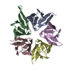

 PDBj
PDBj

 Assembly

 Mass: 18.015 Da / Num. of mol.: 672 / Source method: isolated from a natural source / Formula: H2O
 Sample preparation
 / Beamline: X8C / Wavelength: 0.9795 Å
 Processing