 Movie
Movie Controller

[English] 日本語
 Yorodumi
- PDB-1e4i: 2-deoxy-2-fluoro-beta-D-glucosyl/enzyme intermediate complex of t... -
+ Open data

- Basic information
| Entry | Database: PDB / ID: 1e4i | ||||||
|---|---|---|---|---|---|---|---|
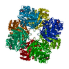
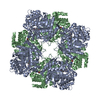
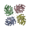
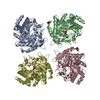
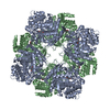
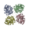
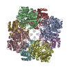
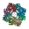
| Title | 2-deoxy-2-fluoro-beta-D-glucosyl/enzyme intermediate complex of the beta-glucosidase from Bacillus polymyxa | ||||||
 Components Components | BETA-GLUCOSIDASE | ||||||
 Keywords Keywords | HYDROLASE / FAMILY 1 GLYCOSYL HYDROLASE / COVALENT ENZYME-GLYCOSIDE INTERMEDIATE / ALPHA/BETA BARREL | ||||||
| Function / homology |  Function and homology information Function and homology informationbeta-glucosidase / beta-glucosidase activity / cellulose catabolic process / cytosol Similarity search - Function | ||||||
| Biological species |  BACILLUS POLYMYXA (bacteria) BACILLUS POLYMYXA (bacteria) | ||||||
| Method |  X-RAY DIFFRACTION / SYNCHROTRON / MOLECULAR REPLACEMENT / Resolution: 2 Å X-RAY DIFFRACTION / SYNCHROTRON / MOLECULAR REPLACEMENT / Resolution: 2 Å | ||||||
 Authors Authors | Sanz-Aparicio, J. / Gonzalez, B. / Hermoso, J.A. / Arribas, J.C. / Canada, F.J. / Polaina, J. | ||||||
 Citation Citation | Journal: Proteins: Struct.,Funct., Genet. / Year: 1998 Title: Structural Basis of Increased Resistance to Thermal Denaturation Induced by Single Amino Acid Substitution in the Sequence of Beta-Glucosidase a from Bacillus Polymyxa. Authors: Sanz-Aparicio, J. / Hermoso, J.A. / Martinez-Ripoll, M. / Gonzalez, B. / Lopez-Camacho, C. / Polaina, J. #1: Journal: J.Mol.Biol. / Year: 1998Title: Crystal Structure of Beta-Glucosidase a from Bacillus Polymyxa: Insights Into the Catalytic Activity in Family 1 Glycosyl Hydrolases Authors: Sanz-Aparicio, J. / Hermoso, J.A. / Martinez-Ripoll, M. / Gonzalez, B. / Lopez-Camacho, C. / Polaina, J. | ||||||
| History |
|
- Structure visualization
| Structure viewer | Molecule: MolmilJmol/JSmol |
|---|
- Downloads & links
-Download
| PDBx/mmCIF format | 1e4i.cif.gz | 108.8 KB | Display | PDBx/mmCIF format |
|---|---|---|---|---|
| PDB format | pdb1e4i.ent.gz | 83.7 KB | Display | PDB format |
| PDBx/mmJSON format | 1e4i.json.gz | Tree view | PDBx/mmJSON format | |
| Others |  Other downloads Other downloads |
-Validation report
| Arichive directory | https://data.pdbj.org/pub/pdb/validation_reports/e4/1e4iftp://data.pdbj.org/pub/pdb/validation_reports/e4/1e4i | HTTPS FTP |
|---|
-Related structure data
| Related structure data |  1bgaS S: Starting model for refinement |
|---|---|
| Similar structure data |


-Links
 PDBj
PDBj- Assembly
| Deposited unit | 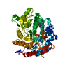
| ||||||||
|---|---|---|---|---|---|---|---|---|---|
| 1 | x 8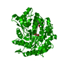
| ||||||||
| Unit cell |
| ||||||||
| Details | BIOMOLECULE |
-Components
| #1: Protein | Mass: 51566.570 Da / Num. of mol.: 1 / Mutation: YES Source method: isolated from a genetically manipulated source Source: (gene. exp.) BACILLUS POLYMYXA (bacteria) / Production host: |
|---|---|
| #2: Sugar | ChemComp-G2F /   Type: D-saccharide, alpha linking / Mass: 182.147 Da / Num. of mol.: 1 Type: D-saccharide, alpha linking / Mass: 182.147 Da / Num. of mol.: 1Source method: isolated from a genetically manipulated source Formula: C6H11FO5 |
| #3: Sugar | ChemComp-NFG /   Type: D-saccharide / Mass: 348.238 Da / Num. of mol.: 1 / Source method: obtained synthetically / Formula: C12H13FN2O9 Type: D-saccharide / Mass: 348.238 Da / Num. of mol.: 1 / Source method: obtained synthetically / Formula: C12H13FN2O9 |
| #4: Water | ChemComp-HOH /  Mass: 18.015 Da / Num. of mol.: 234 / Source method: isolated from a natural source / Formula: H2O Mass: 18.015 Da / Num. of mol.: 234 / Source method: isolated from a natural source / Formula: H2O |
| Has protein modification | Y |
-Experimental details
-Experiment
| Experiment | Method: X-RAY DIFFRACTION / Number of used crystals: 1 |
|---|
- Sample preparation
| Crystal | Density Matthews: 4.1 Å3/Da / Density % sol: 70 % |
|---|---|
| Crystal grow | pH: 7 Details: DROP: 10 MICROL PROTEIN (8 MG/ML)+ 5 MICROL PP(1M, PH=7)+5 MICROL INH (5MM) RESERVOIR: 1.3M PP, pH 7.00 |
-Data collection
| Diffraction | Mean temperature: 100 K |
|---|---|
| Diffraction source | Source: SYNCHROTRON / Site: ESRF  / Beamline: BM02 / Wavelength: 0.989 / Beamline: BM02 / Wavelength: 0.989 |
| Detector | Detector: CCD / Date: Oct 15, 1996 |
| Radiation | Protocol: SINGLE WAVELENGTH / Monochromatic (M) / Laue (L): M / Scattering type: x-ray |
| Radiation wavelength | Wavelength: 0.989 Å / Relative weight: 1 |
| Reflection | Resolution: 1.96→42.3 Å / Num. obs: 56246 / % possible obs: 98 % / Redundancy: 5 % / Rmerge(I) obs: 0.04 |
| Reflection shell | Resolution: 2→2.1 Å / Rsym value: 0.34 / % possible all: 95 |
- Processing
| Software |
| ||||||||||||||||||||||||||||||||||||||||||||||||||||||||||||
|---|---|---|---|---|---|---|---|---|---|---|---|---|---|---|---|---|---|---|---|---|---|---|---|---|---|---|---|---|---|---|---|---|---|---|---|---|---|---|---|---|---|---|---|---|---|---|---|---|---|---|---|---|---|---|---|---|---|---|---|---|---|
| Refinement | Method to determine structure: MOLECULAR REPLACEMENT Starting model: PDB ENTRY 1BGA Resolution: 2→8 Å / Data cutoff high absF: 100000 / Data cutoff low absF: 0.1 / Isotropic thermal model: RESTRAINED / Cross valid method: THROUGHOUT / σ(F): 0
| ||||||||||||||||||||||||||||||||||||||||||||||||||||||||||||
| Displacement parameters | Biso mean: 15.9 Å2 | ||||||||||||||||||||||||||||||||||||||||||||||||||||||||||||
| Refinement step | Cycle: LAST / Resolution: 2→8 Å
| ||||||||||||||||||||||||||||||||||||||||||||||||||||||||||||
| Refine LS restraints |
| ||||||||||||||||||||||||||||||||||||||||||||||||||||||||||||
| LS refinement shell | Resolution: 2→2.09 Å / Total num. of bins used: 8 /
| ||||||||||||||||||||||||||||||||||||||||||||||||||||||||||||
| Xplor file |
|
