 Movie
Movie Controller

[English] 日本語
 Yorodumi
- PDB-6r4k: Structure of beta-glucosidase A from Paenibacillus polymyxa compl... -
+ Open data

- Basic information
| Entry | Database: PDB / ID: 6r4k | ||||||
|---|---|---|---|---|---|---|---|
| Title | Structure of beta-glucosidase A from Paenibacillus polymyxa complexed with a monovalent inhibitor | ||||||
 Components Components | Beta-glucosidase A | ||||||
 Keywords Keywords | HYDROLASE / BETA-GLUCOSIDASE / GLYCOSIDASE / CARBOHYDRATE / CARBOHYDRATE METABOLISM / POLISACCHARIDE DEGRADATION / COMPLEX / INHIBITOR | ||||||
| Function / homology |  Function and homology information Function and homology informationbeta-glucosidase / beta-glucosidase activity / cellulose catabolic process / cytosol Similarity search - Function | ||||||
| Biological species |  Paenibacillus polymyxa (bacteria) Paenibacillus polymyxa (bacteria) | ||||||
| Method |  X-RAY DIFFRACTION / SYNCHROTRON / MOLECULAR REPLACEMENT / Resolution: 2.13 Å X-RAY DIFFRACTION / SYNCHROTRON / MOLECULAR REPLACEMENT / Resolution: 2.13 Å | ||||||
 Authors Authors | Jimenez-Ortega, E. / Sanz-Aparicio, J. | ||||||
 Citation Citation | Journal: Bioorg.Chem. / Year: 2019 Title: Structural basis of the inhibition of GH1 beta-glucosidases by multivalent pyrrolidine iminosugars. Authors: Martinez-Bailen, M. / Jimenez-Ortega, E. / Carmona, A.T. / Robina, I. / Sanz-Aparicio, J. / Talens-Perales, D. / Polaina, J. / Matassini, C. / Cardona, F. / Moreno-Vargas, A.J. | ||||||
| History |
|
- Structure visualization
| Structure viewer | Molecule: MolmilJmol/JSmol |
|---|
- Downloads & links
-Download
| PDBx/mmCIF format | 6r4k.cif.gz | 383.9 KB | Display | PDBx/mmCIF format |
|---|---|---|---|---|
| PDB format | pdb6r4k.ent.gz | 317.1 KB | Display | PDB format |
| PDBx/mmJSON format | 6r4k.json.gz | Tree view | PDBx/mmJSON format | |
| Others |  Other downloads Other downloads |
-Validation report
| Arichive directory | https://data.pdbj.org/pub/pdb/validation_reports/r4/6r4kftp://data.pdbj.org/pub/pdb/validation_reports/r4/6r4k | HTTPS FTP |
|---|
-Related structure data
| Related structure data |  6qwiC  1e4iS S: Starting model for refinement C: citing same article ( |
|---|---|
| Similar structure data |
-Links
 PDBj
PDBj- Assembly
| Deposited unit | 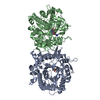
| ||||||||
|---|---|---|---|---|---|---|---|---|---|
| 1 | 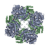
| ||||||||
| Unit cell |
|
-Components
| #1: Protein | Mass: 51608.719 Da / Num. of mol.: 2 Source method: isolated from a genetically manipulated source Source: (gene. exp.) Paenibacillus polymyxa (bacteria) / Gene: bglA / Plasmid: pQE-80L / Production host: #2: Chemical |   Mass: 391.425 Da / Num. of mol.: 2 / Source method: obtained synthetically / Formula: C17H25N7O4 Mass: 391.425 Da / Num. of mol.: 2 / Source method: obtained synthetically / Formula: C17H25N7O4#3: Water | ChemComp-HOH / |  Mass: 18.015 Da / Num. of mol.: 458 / Source method: isolated from a natural source / Formula: H2O Mass: 18.015 Da / Num. of mol.: 458 / Source method: isolated from a natural source / Formula: H2O |
|---|
-Experimental details
-Experiment
| Experiment | Method: X-RAY DIFFRACTION / Number of used crystals: 1 |
|---|
- Sample preparation
| Crystal | Density Matthews: 3.65 Å3/Da / Density % sol: 66.31 % / Description: Prismatic crystals |
|---|---|
| Crystal grow | Temperature: 291 K / Method: vapor diffusion, sitting drop / pH: 7.5 Details: 18% (v/v) PEG 3350, 0.2M sodium nitrate, 3% 2-Methyl-2,4-pentanediol (MPD), 0.1 M Bis-Tris propane, pH 7.5, and 5% n-Dodecyl beta-D-maltoside. Cryoprotectant mother liquor supplemented with 25% glycerol. |
-Data collection
| Diffraction | Mean temperature: 100 K / Serial crystal experiment: N |
|---|---|
| Diffraction source | Source: SYNCHROTRON / Site: ALBA  / Beamline: XALOC / Wavelength: 0.97926 Å / Beamline: XALOC / Wavelength: 0.97926 Å |
| Detector | Type: DECTRIS PILATUS3 S 6M / Detector: PIXEL / Date: Jul 22, 2018 / Details: KB focusing mirrors |
| Radiation | Monochromator: Si(111) channel-cut, cryocooled / Protocol: SINGLE WAVELENGTH / Monochromatic (M) / Laue (L): M / Scattering type: x-ray |
| Radiation wavelength | Wavelength: 0.97926 Å / Relative weight: 1 |
| Reflection | Resolution: 2.13→48.91 Å / Num. obs: 85545 / % possible obs: 99.9 % / Redundancy: 6.7 % / CC1/2: 0.991 / Rmerge(I) obs: 0.104 / Rpim(I) all: 0.043 / Rrim(I) all: 0.113 / Net I/σ(I): 11.6 |
| Reflection shell | Resolution: 2.13→2.17 Å / Redundancy: 6.2 % / Rmerge(I) obs: 0.701 / Mean I/σ(I) obs: 2.2 / Num. unique obs: 4461 / CC1/2: 0.655 / Rpim(I) all: 0.299 / Rrim(I) all: 0.765 / % possible all: 99.6 |
- Processing
| Software |
| |||||||||||||||||||||||||||||||||||||||||||||||||||||||||||||||||||||||||||||||||||||||||||||||||||||||||||||||||||||||||||||||||||||||||||||||||||
|---|---|---|---|---|---|---|---|---|---|---|---|---|---|---|---|---|---|---|---|---|---|---|---|---|---|---|---|---|---|---|---|---|---|---|---|---|---|---|---|---|---|---|---|---|---|---|---|---|---|---|---|---|---|---|---|---|---|---|---|---|---|---|---|---|---|---|---|---|---|---|---|---|---|---|---|---|---|---|---|---|---|---|---|---|---|---|---|---|---|---|---|---|---|---|---|---|---|---|---|---|---|---|---|---|---|---|---|---|---|---|---|---|---|---|---|---|---|---|---|---|---|---|---|---|---|---|---|---|---|---|---|---|---|---|---|---|---|---|---|---|---|---|---|---|---|---|---|---|
| Refinement | Method to determine structure: MOLECULAR REPLACEMENT Starting model: 1E4I Resolution: 2.13→48.646 Å / Cross valid method: FREE R-VALUE / σ(F): 83 / Phase error: 28.92
| |||||||||||||||||||||||||||||||||||||||||||||||||||||||||||||||||||||||||||||||||||||||||||||||||||||||||||||||||||||||||||||||||||||||||||||||||||
| Solvent computation | Shrinkage radii: 0.9 Å / VDW probe radii: 1.11 Å | |||||||||||||||||||||||||||||||||||||||||||||||||||||||||||||||||||||||||||||||||||||||||||||||||||||||||||||||||||||||||||||||||||||||||||||||||||
| Refinement step | Cycle: LAST / Resolution: 2.13→48.646 Å
| |||||||||||||||||||||||||||||||||||||||||||||||||||||||||||||||||||||||||||||||||||||||||||||||||||||||||||||||||||||||||||||||||||||||||||||||||||
| Refine LS restraints |
| |||||||||||||||||||||||||||||||||||||||||||||||||||||||||||||||||||||||||||||||||||||||||||||||||||||||||||||||||||||||||||||||||||||||||||||||||||
| LS refinement shell |
| |||||||||||||||||||||||||||||||||||||||||||||||||||||||||||||||||||||||||||||||||||||||||||||||||||||||||||||||||||||||||||||||||||||||||||||||||||
| Refinement TLS params. | Method: refined / Origin x: 11.8397 Å / Origin y: -39.7753 Å / Origin z: 45.1114 Å
| |||||||||||||||||||||||||||||||||||||||||||||||||||||||||||||||||||||||||||||||||||||||||||||||||||||||||||||||||||||||||||||||||||||||||||||||||||
| Refinement TLS group | Selection details: all |
