 Movie
Movie Controller


+ Open data

- Basic information
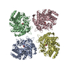
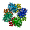
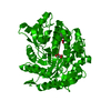
| Entry | Database: PDB / ID: 1qox | ||||||
|---|---|---|---|---|---|---|---|
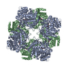
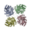
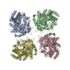
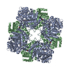
| Title | Beta-glucosidase from Bacillus circulans sp. alkalophilus | ||||||
 Components Components | BETA-GLUCOSIDASE | ||||||
 Keywords Keywords | HYDROLASE / CELLULOSE DEGRADATION | ||||||
| Function / homology |  Function and homology information Function and homology informationbeta-glucosidase / beta-glucosidase activity / cellulose catabolic process Similarity search - Function | ||||||
| Biological species |  BACILLUS CIRCULANS (bacteria) BACILLUS CIRCULANS (bacteria) | ||||||
| Method |  X-RAY DIFFRACTION / MOLECULAR REPLACEMENT / Resolution: 2.7 Å X-RAY DIFFRACTION / MOLECULAR REPLACEMENT / Resolution: 2.7 Å | ||||||
 Authors Authors | Hakulinen, N. / Rouvinen, J. | ||||||
 Citation Citation | Journal: J.Struct.Biol. / Year: 2000 Title: The Crystal Structure of Beta-Glucosidase from Bacillus Circulans Sp. Alkalophilus: Ability to Form Long Polymeric Assemblies Authors: Hakulinen, N. / Paavilainen, S. / Korpela, T. / Rouvinen, J. | ||||||
| History |
| ||||||
| Remark 700 | SHEET DETERMINATION METHOD: DSSP THE SHEETS PRESENTED AS "B" IN EACH CHAIN (E.G. SHEET AB IN CHAIN ... SHEET DETERMINATION METHOD: DSSP THE SHEETS PRESENTED AS "B" IN EACH CHAIN (E.G. SHEET AB IN CHAIN A) ON SHEET RECORDS BELOW IS ACTUALLY AN 8-STRANDED BETA-BARREL. THIS IS REPRESENTED BY A 9-STRANDED SHEET IN WHICH THE FIRST AND LAST STRANDS ARE IDENTICAL. |
- Structure visualization
| Structure viewer | Molecule: MolmilJmol/JSmol |
|---|
- Downloads & links
-Download
| PDBx/mmCIF format | 1qox.cif.gz | 1.4 MB | Display | PDBx/mmCIF format |
|---|---|---|---|---|
| PDB format | pdb1qox.ent.gz | 1.2 MB | Display | PDB format |
| PDBx/mmJSON format | 1qox.json.gz | Tree view | PDBx/mmJSON format | |
| Others |  Other downloads Other downloads |
-Validation report
| Arichive directory | https://data.pdbj.org/pub/pdb/validation_reports/qo/1qoxftp://data.pdbj.org/pub/pdb/validation_reports/qo/1qox | HTTPS FTP |
|---|
-Related structure data
| Related structure data |  1bgaS S: Starting model for refinement |
|---|---|
| Similar structure data |

-Links
 PDBj
PDBj- Assembly
| Deposited unit | 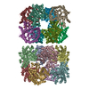
| ||||||||||||||||||||||||||||||||||||||||||||||||||||||||||||||||
|---|---|---|---|---|---|---|---|---|---|---|---|---|---|---|---|---|---|---|---|---|---|---|---|---|---|---|---|---|---|---|---|---|---|---|---|---|---|---|---|---|---|---|---|---|---|---|---|---|---|---|---|---|---|---|---|---|---|---|---|---|---|---|---|---|---|
| 1 | 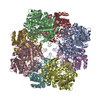
| ||||||||||||||||||||||||||||||||||||||||||||||||||||||||||||||||
| 2 | 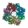
| ||||||||||||||||||||||||||||||||||||||||||||||||||||||||||||||||
| Unit cell |
| ||||||||||||||||||||||||||||||||||||||||||||||||||||||||||||||||
| Noncrystallographic symmetry (NCS) | NCS oper:
|
-Components
| #1: Protein | Mass: 51222.477 Da / Num. of mol.: 16 / Source method: isolated from a natural source / Source: (natural) BACILLUS CIRCULANS (bacteria) / Strain: ALKALOPHILUS / References: UniProt: Q03506, beta-glucosidase#2: Water | ChemComp-HOH / |  Mass: 18.015 Da / Num. of mol.: 2448 / Source method: isolated from a natural source / Formula: H2O Mass: 18.015 Da / Num. of mol.: 2448 / Source method: isolated from a natural source / Formula: H2O |
|---|
-Experimental details
-Experiment
| Experiment | Method: X-RAY DIFFRACTION / Number of used crystals: 1 |
|---|
- Sample preparation
| Crystal | Density Matthews: 2.5 Å3/Da / Density % sol: 51 % | ||||||||||||||||||||||||||||||
|---|---|---|---|---|---|---|---|---|---|---|---|---|---|---|---|---|---|---|---|---|---|---|---|---|---|---|---|---|---|---|---|
| Crystal grow | pH: 6 Details: 13 % PEG8000, 0.3 M SODIUM FORMATE, 0.1 M SODIUM ACETATE, PH 6.0 | ||||||||||||||||||||||||||||||
| Crystal grow | *PLUS pH: 7.5 / Method: vapor diffusion, hanging drop | ||||||||||||||||||||||||||||||
| Components of the solutions | *PLUS
|
-Data collection
| Diffraction | Mean temperature: 100 K |
|---|---|
| Diffraction source | Source: ROTATING ANODE / Type: RIGAKU RUH2R / Wavelength: 1.5418 |
| Detector | Type: RIGAKU IMAGE PLATE / Detector: IMAGE PLATE / Date: Aug 15, 1997 |
| Radiation | Monochromator: GRAPHITE(002) / Protocol: SINGLE WAVELENGTH / Monochromatic (M) / Laue (L): M / Scattering type: x-ray |
| Radiation wavelength | Wavelength: 1.5418 Å / Relative weight: 1 |
| Reflection | Resolution: 2.69→99 Å / Num. obs: 186165 / % possible obs: 83.7 % / Redundancy: 1.63 % / Biso Wilson estimate: 51 Å2 / Rsym value: 0.076 / Net I/σ(I): 8.7 |
| Reflection shell | Resolution: 2.69→2.78 Å / Redundancy: 1.47 % / Mean I/σ(I) obs: 1.7 / Rsym value: 0.322 / % possible all: 66 |
| Reflection | *PLUS Num. measured all: 303163 / Rmerge(I) obs: 0.076 |
| Reflection shell | *PLUS % possible obs: 66 % / Rmerge(I) obs: 0.322 |
- Processing
| Software |
| ||||||||||||||||||||||||||||||||||||||||||||||||||||||||||||
|---|---|---|---|---|---|---|---|---|---|---|---|---|---|---|---|---|---|---|---|---|---|---|---|---|---|---|---|---|---|---|---|---|---|---|---|---|---|---|---|---|---|---|---|---|---|---|---|---|---|---|---|---|---|---|---|---|---|---|---|---|---|
| Refinement | Method to determine structure: MOLECULAR REPLACEMENT Starting model: 1BGA Resolution: 2.7→10 Å / Data cutoff high absF: 100000 / Data cutoff low absF: 0.1 / σ(F): 0
| ||||||||||||||||||||||||||||||||||||||||||||||||||||||||||||
| Displacement parameters | Biso mean: 21.1 Å2 | ||||||||||||||||||||||||||||||||||||||||||||||||||||||||||||
| Refinement step | Cycle: LAST / Resolution: 2.7→10 Å
| ||||||||||||||||||||||||||||||||||||||||||||||||||||||||||||
| Refine LS restraints |
| ||||||||||||||||||||||||||||||||||||||||||||||||||||||||||||
| Xplor file | Serial no: 1 / Param file: PARHCSDX.PRO / Topol file: TOPHCSDX.PRO | ||||||||||||||||||||||||||||||||||||||||||||||||||||||||||||
| Software | *PLUS Name: X-PLOR / Version: 3.851 / Classification: refinement | ||||||||||||||||||||||||||||||||||||||||||||||||||||||||||||
| Refine LS restraints | *PLUS
|
