 Movie
Movie Controller


+ Open data

- Basic information
| Entry | Database: PDB / ID: 1bgg | |||||||||
|---|---|---|---|---|---|---|---|---|---|---|
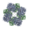
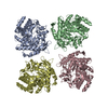
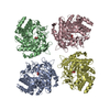
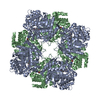
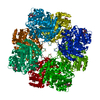
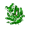
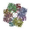
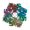
| Title | GLUCOSIDASE A FROM BACILLUS POLYMYXA COMPLEXED WITH GLUCONATE | |||||||||
 Components Components | BETA-GLUCOSIDASE A | |||||||||
 Keywords Keywords | FAMILY 1 BETA-GLUCOSIDASE COMPLEX / GLYCOSYL-HYDROLASE COMPLEX | |||||||||
| Function / homology |  Function and homology information Function and homology informationbeta-glucosidase / beta-glucosidase activity / cellulose catabolic process / cytosol Similarity search - Function | |||||||||
| Biological species |  Paenibacillus polymyxa (bacteria) Paenibacillus polymyxa (bacteria) | |||||||||
| Method |  X-RAY DIFFRACTION / SYNCHROTRON / DIFFERENCE FOURIER / Resolution: 2.3 Å X-RAY DIFFRACTION / SYNCHROTRON / DIFFERENCE FOURIER / Resolution: 2.3 Å | |||||||||
 Authors Authors | Sanz-Aparicio, J. / Hermoso, J. / Martinez-Ripoll, M. / Polaina, J. | |||||||||
 Citation Citation | Journal: J.Mol.Biol. / Year: 1998 Title: Crystal structure of beta-glucosidase A from Bacillus polymyxa: insights into the catalytic activity in family 1 glycosyl hydrolases. Authors: Sanz-Aparicio, J. / Hermoso, J.A. / Martinez-Ripoll, M. / Lequerica, J.L. / Polaina, J. #1: Journal: J.Mol.Biol. / Year: 1994Title: Crystallization and Preliminary X-Ray Diffraction Analysis of a Type I Beta-Glucosidase Encoded by the Bgia Gene of Bacillus Polymyxa Authors: Sanz-Aparicio, J. / Romero, A. / Martinez-Ripoll, M. / Madarro, A. / Flors, A. / Polaina, J. #2: Journal: J.Bacteriol. / Year: 1992Title: Purification and Characterization of a Bacillus Polymyxa Beta-Glucosidase Expressed in Escherichia Coli Authors: Painbeni, E. / Valles, S. / Polaina, J. / Flors, A. | |||||||||
| History |
|
- Structure visualization
| Structure viewer | Molecule: MolmilJmol/JSmol |
|---|
- Downloads & links
-Download
| PDBx/mmCIF format | 1bgg.cif.gz | 391.1 KB | Display | PDBx/mmCIF format |
|---|---|---|---|---|
| PDB format | pdb1bgg.ent.gz | 318.9 KB | Display | PDB format |
| PDBx/mmJSON format | 1bgg.json.gz | Tree view | PDBx/mmJSON format | |
| Others |  Other downloads Other downloads |
-Validation report
| Arichive directory | https://data.pdbj.org/pub/pdb/validation_reports/bg/1bggftp://data.pdbj.org/pub/pdb/validation_reports/bg/1bgg | HTTPS FTP |
|---|
-Related structure data
| Related structure data |  1bgaSC  1tr1C S: Starting model for refinement C: citing same article ( |
|---|---|
| Similar structure data |
-Links
 PDBj
PDBj- Assembly
| Deposited unit | 
| ||||||||||||||||
|---|---|---|---|---|---|---|---|---|---|---|---|---|---|---|---|---|---|
| 1 | 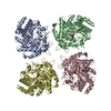
| ||||||||||||||||
| Unit cell |
| ||||||||||||||||
| Noncrystallographic symmetry (NCS) | NCS oper:
|
-Components
| #1: Protein | Mass: 51698.688 Da / Num. of mol.: 4 Source method: isolated from a genetically manipulated source Source: (gene. exp.) Paenibacillus polymyxa (bacteria) / Gene: BGLA / Plasmid: PUC DERIVATIVE / Production host: #2: Sugar |   Type: D-saccharide / Mass: 196.155 Da / Num. of mol.: 3 Type: D-saccharide / Mass: 196.155 Da / Num. of mol.: 3Source method: isolated from a genetically manipulated source Formula: C6H12O7 #3: Water | ChemComp-HOH / |  Mass: 18.015 Da / Num. of mol.: 1494 / Source method: isolated from a natural source / Formula: H2O Mass: 18.015 Da / Num. of mol.: 1494 / Source method: isolated from a natural source / Formula: H2O |
|---|
-Experimental details
-Experiment
| Experiment | Method: X-RAY DIFFRACTION / Number of used crystals: 1 |
|---|
- Sample preparation
| Crystal | Density Matthews: 4.2 Å3/Da / Density % sol: 70 % | |||||||||||||||
|---|---|---|---|---|---|---|---|---|---|---|---|---|---|---|---|---|
| Crystal grow | Method: co-crystallization / pH: 8.3 Details: COMPLEX WAS OBTAINED BY CO-CRYSTALLIZATION, 5 MICRO-L BGLA (14 MG/ML) / 5 MICRO-L 10MM LIGAND / 5 MICRO-L PP 1.3M, PH 8.3, co-crystallization | |||||||||||||||
| Crystal grow | *PLUS Method: co-crystallization | |||||||||||||||
| Components of the solutions | *PLUS
|
-Data collection
| Diffraction | Mean temperature: 176 K |
|---|---|
| Diffraction source | Source: SYNCHROTRON / Site: LURE  / Type: LURE / Wavelength: 0.983 / Type: LURE / Wavelength: 0.983 |
| Detector | Type: MARRESEARCH / Detector: IMAGE PLATE / Date: Oct 1, 1996 |
| Radiation | Monochromatic (M) / Laue (L): M / Scattering type: x-ray |
| Radiation wavelength | Wavelength: 0.983 Å / Relative weight: 1 |
| Reflection | Resolution: 2.3→31.4 Å / Num. obs: 146539 / % possible obs: 99.4 % / Observed criterion σ(I): 4 / Redundancy: 5.7 % / Rmerge(I) obs: 0.09 / Rsym value: 0.09 / Net I/σ(I): 6.2 |
| Reflection shell | Resolution: 2.3→2.36 Å / Redundancy: 3.8 % / Rmerge(I) obs: 0.32 / Mean I/σ(I) obs: 2 / Rsym value: 0.32 / % possible all: 97.2 |
| Reflection | *PLUS Num. measured all: 840221 / Rmerge(I) obs: 0.092 |
| Reflection shell | *PLUS % possible obs: 97.2 % |
- Processing
| Software |
| ||||||||||||||||||||||||||||||||||||||||||||||||||||||||||||
|---|---|---|---|---|---|---|---|---|---|---|---|---|---|---|---|---|---|---|---|---|---|---|---|---|---|---|---|---|---|---|---|---|---|---|---|---|---|---|---|---|---|---|---|---|---|---|---|---|---|---|---|---|---|---|---|---|---|---|---|---|---|
| Refinement | Method to determine structure: DIFFERENCE FOURIER Starting model: PDB ENTRY 1BGA Resolution: 2.3→8 Å / Data cutoff high absF: 1000000 / Data cutoff low absF: 0.001 / Cross valid method: THROUGHOUT / σ(F): 2
| ||||||||||||||||||||||||||||||||||||||||||||||||||||||||||||
| Displacement parameters | Biso mean: 14.4 Å2 | ||||||||||||||||||||||||||||||||||||||||||||||||||||||||||||
| Refinement step | Cycle: LAST / Resolution: 2.3→8 Å
| ||||||||||||||||||||||||||||||||||||||||||||||||||||||||||||
| Refine LS restraints |
| ||||||||||||||||||||||||||||||||||||||||||||||||||||||||||||
| LS refinement shell | Resolution: 2.3→2.4 Å / Total num. of bins used: 8
| ||||||||||||||||||||||||||||||||||||||||||||||||||||||||||||
| Software | *PLUS Name: X-PLOR / Version: 3.843 / Classification: refinement | ||||||||||||||||||||||||||||||||||||||||||||||||||||||||||||
| Refinement | *PLUS Num. reflection obs: 14576 | ||||||||||||||||||||||||||||||||||||||||||||||||||||||||||||
| Solvent computation | *PLUS | ||||||||||||||||||||||||||||||||||||||||||||||||||||||||||||
| Displacement parameters | *PLUS | ||||||||||||||||||||||||||||||||||||||||||||||||||||||||||||
| Refine LS restraints | *PLUS
|
