 Movie
Movie Controller

[English] 日本語
 Yorodumi
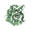
- PDB-3ahx: Crystal structure of beta-glucosidase A from bacterium Clostridiu... -
+ Open data

- Basic information
| Entry | Database: PDB / ID: 3ahx | ||||||
|---|---|---|---|---|---|---|---|
| Title | Crystal structure of beta-glucosidase A from bacterium Clostridium cellulovorans | ||||||
 Components Components | Beta-glucosidase A | ||||||
 Keywords Keywords | HYDROLASE / cellulases / glycosyl hydrolase / manganese enhancement | ||||||
| Function / homology |  Function and homology information Function and homology informationbeta-glucosidase / beta-glucosidase activity / cellulose catabolic process / cytosol Similarity search - Function | ||||||
| Biological species |  Clostridium cellulovorans (bacteria) Clostridium cellulovorans (bacteria) | ||||||
| Method |  X-RAY DIFFRACTION / SYNCHROTRON / MOLECULAR REPLACEMENT / Resolution: 1.9 Å X-RAY DIFFRACTION / SYNCHROTRON / MOLECULAR REPLACEMENT / Resolution: 1.9 Å | ||||||
 Authors Authors | Jeng, W.-Y. / Liu, C.-I. / Wang, A.H.-J. | ||||||
 Citation Citation | Journal: J.Struct.Biol. / Year: 2011 Title: Structural and functional analysis of three beta-glucosidases from bacterium Clostridium cellulovorans, fungus Trichoderma reesei and termite Neotermes koshunensis Authors: Jeng, W.-Y. / Wang, N.-C. / Lin, M.-H. / Lin, C.-T. / Liaw, Y.-C. / Chang, W.-J. / Liu, C.-I. / Liang, P.-H. / Wang, A.H.-J. | ||||||
| History |
|
- Structure visualization
| Structure viewer | Molecule: MolmilJmol/JSmol |
|---|
- Downloads & links
-Download
| PDBx/mmCIF format | 3ahx.cif.gz | 785.4 KB | Display | PDBx/mmCIF format |
|---|---|---|---|---|
| PDB format | pdb3ahx.ent.gz | 652 KB | Display | PDB format |
| PDBx/mmJSON format | 3ahx.json.gz | Tree view | PDBx/mmJSON format | |
| Others |  Other downloads Other downloads |
-Validation report
| Arichive directory | https://data.pdbj.org/pub/pdb/validation_reports/ah/3ahxftp://data.pdbj.org/pub/pdb/validation_reports/ah/3ahx | HTTPS FTP |
|---|
-Related structure data
| Related structure data |  3ahyC  3ahzC  3ai0C  1od0S C: citing same article ( S: Starting model for refinement |
|---|---|
| Similar structure data |


-Links
 PDBj
PDBj- Assembly
| Deposited unit | 
| ||||||||
|---|---|---|---|---|---|---|---|---|---|
| 1 | 
| ||||||||
| 2 | 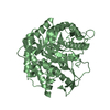
| ||||||||
| 3 | 
| ||||||||
| 4 | 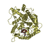
| ||||||||
| Unit cell |
|
-Components
| #1: Protein | Mass: 52698.105 Da / Num. of mol.: 4 Source method: isolated from a genetically manipulated source Source: (gene. exp.) Clostridium cellulovorans (bacteria) / Gene: BglA / Plasmid: pET-21a / Production host: #2: Chemical |   Mass: 310.384 Da / Num. of mol.: 2 / Source method: obtained synthetically / Formula: C14H30O7 Mass: 310.384 Da / Num. of mol.: 2 / Source method: obtained synthetically / Formula: C14H30O7#3: Water | ChemComp-HOH / |  Mass: 18.015 Da / Num. of mol.: 2147 / Source method: isolated from a natural source / Formula: H2O Mass: 18.015 Da / Num. of mol.: 2147 / Source method: isolated from a natural source / Formula: H2O |
|---|
-Experimental details
-Experiment
| Experiment | Method: X-RAY DIFFRACTION / Number of used crystals: 1 |
|---|
- Sample preparation
| Crystal | Density Matthews: 2.59 Å3/Da / Density % sol: 52.43 % |
|---|---|
| Crystal grow | Temperature: 298 K / Method: vapor diffusion, sitting drop / pH: 7.5 Details: 0.1M Hepes, 21-23%(w/v) PEG 3350, 0.3-0.45M Li2SO4, pH 7.5, VAPOR DIFFUSION, SITTING DROP, temperature 298K |
-Data collection
| Diffraction | Mean temperature: 100 K |
|---|---|
| Diffraction source | Source: SYNCHROTRON / Site: NSRRC  / Beamline: BL13B1 / Wavelength: 1 Å / Beamline: BL13B1 / Wavelength: 1 Å |
| Detector | Type: ADSC QUANTUM 315 / Detector: CCD / Date: Nov 9, 2008 Details: Vertically Collimating Premirror, Toroidal Focusing Mirror |
| Radiation | Monochromator: LN2-Cooled Fixed-Exit Double Crystal Si(111) Monochromator Protocol: SINGLE WAVELENGTH / Monochromatic (M) / Laue (L): M / Scattering type: x-ray |
| Radiation wavelength | Wavelength: 1 Å / Relative weight: 1 |
| Reflection | Resolution: 1.9→30 Å / Num. all: 172709 / Num. obs: 171402 / % possible obs: 99.2 % / Observed criterion σ(F): 0 / Observed criterion σ(I): 1 / Redundancy: 6.7 % / Biso Wilson estimate: 31 Å2 / Rmerge(I) obs: 0.062 / Net I/σ(I): 27.5 |
| Reflection shell | Resolution: 1.9→1.97 Å / Redundancy: 3.8 % / Rmerge(I) obs: 0.323 / Mean I/σ(I) obs: 3.8 / Num. unique all: 17061 / % possible all: 98 |
- Processing
| Software |
| |||||||||||||||||||||||||||||||||||||||||||||||||||||||||||||||||||||||||||||||||||||||||||||||||||||||||||||||||||||||||||||
|---|---|---|---|---|---|---|---|---|---|---|---|---|---|---|---|---|---|---|---|---|---|---|---|---|---|---|---|---|---|---|---|---|---|---|---|---|---|---|---|---|---|---|---|---|---|---|---|---|---|---|---|---|---|---|---|---|---|---|---|---|---|---|---|---|---|---|---|---|---|---|---|---|---|---|---|---|---|---|---|---|---|---|---|---|---|---|---|---|---|---|---|---|---|---|---|---|---|---|---|---|---|---|---|---|---|---|---|---|---|---|---|---|---|---|---|---|---|---|---|---|---|---|---|---|---|---|
| Refinement | Method to determine structure: MOLECULAR REPLACEMENT Starting model: PDB ENTRY 1OD0 Resolution: 1.9→28.3 Å / Cor.coef. Fo:Fc: 0.968 / Cor.coef. Fo:Fc free: 0.947 / SU B: 5.954 / SU ML: 0.079 / Isotropic thermal model: Isotropic with TLS / Cross valid method: THROUGHOUT / ESU R: 3.573 / ESU R Free: 0.131 / Stereochemistry target values: MAXIMUM LIKELIHOOD / Details: HYDROGENS HAVE BEEN ADDED IN THE RIDING POSITIONS
| |||||||||||||||||||||||||||||||||||||||||||||||||||||||||||||||||||||||||||||||||||||||||||||||||||||||||||||||||||||||||||||
| Solvent computation | Ion probe radii: 0.8 Å / Shrinkage radii: 0.8 Å / VDW probe radii: 1.4 Å / Solvent model: MASK | |||||||||||||||||||||||||||||||||||||||||||||||||||||||||||||||||||||||||||||||||||||||||||||||||||||||||||||||||||||||||||||
| Displacement parameters | Biso mean: 20.123 Å2
| |||||||||||||||||||||||||||||||||||||||||||||||||||||||||||||||||||||||||||||||||||||||||||||||||||||||||||||||||||||||||||||
| Refinement step | Cycle: LAST / Resolution: 1.9→28.3 Å
| |||||||||||||||||||||||||||||||||||||||||||||||||||||||||||||||||||||||||||||||||||||||||||||||||||||||||||||||||||||||||||||
| Refine LS restraints |
| |||||||||||||||||||||||||||||||||||||||||||||||||||||||||||||||||||||||||||||||||||||||||||||||||||||||||||||||||||||||||||||
| LS refinement shell | Resolution: 1.902→2.005 Å / Total num. of bins used: 10
| |||||||||||||||||||||||||||||||||||||||||||||||||||||||||||||||||||||||||||||||||||||||||||||||||||||||||||||||||||||||||||||
| Refinement TLS params. | Method: refined / Refine-ID: X-RAY DIFFRACTION
| |||||||||||||||||||||||||||||||||||||||||||||||||||||||||||||||||||||||||||||||||||||||||||||||||||||||||||||||||||||||||||||
| Refinement TLS group |
|
