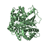
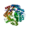
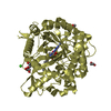
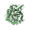
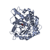
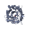
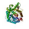
- PDB-5idi: Structure of beta glucosidase 1A from Thermotoga neapolitana, mut... -
+
Open data
ID or keywords:
Loading...
-
Basic information
Entry
Database: PDB / ID: 5idi
Title
Structure of beta glucosidase 1A from Thermotoga neapolitana, mutant E349A
Components
1,4-beta-D-glucan glucohydrolase
Keywords
HYDROLASE / beta-glucosidase / glycosyl hydrolase family 1
Function / homology
Function and homology information
glucan 1,4-beta-glucosidase / glucan 1,4-beta-glucosidase activity / beta-glucosidase / cellulose catabolic process / cytosol Similarity search - Function
Glycoside hydrolase, family 1, beta-glucosidase / Glycoside hydrolase family 1, active site / Glycosyl hydrolases family 1 active site. / Glycosyl hydrolases family 1, N-terminal conserved site / Glycosyl hydrolases family 1 N-terminal signature. / Glycosyl hydrolase family 1 / Glycoside hydrolase family 1 / Glycosidases / Glycoside hydrolase superfamily / TIM Barrel ...Glycoside hydrolase, family 1, beta-glucosidase / Glycoside hydrolase family 1, active site / Glycosyl hydrolases family 1 active site. / Glycosyl hydrolases family 1, N-terminal conserved site / Glycosyl hydrolases family 1 N-terminal signature. / Glycosyl hydrolase family 1 / Glycoside hydrolase family 1 / Glycosidases / Glycoside hydrolase superfamily / TIM Barrel / Alpha-Beta Barrel / Alpha Beta Similarity search - Domain/homology
Mass: 18.015 Da / Num. of mol.: 429 / Source method: isolated from a natural source / Formula: H2O
-
Experimental details
-
Experiment
Experiment
Method: X-RAY DIFFRACTION / Number of used crystals: 1
-
Sample preparation
Crystal
Density Matthews: 2.47 Å3/Da / Density % sol: 50.15 %
Crystal grow
Temperature: 288 K / Method: vapor diffusion / pH: 5 Details: Protein at 12 mg/ml in 20 mM citrate phosphate buffer, pH 5.6. Hanging drops consisting of 1 microlitre of protein solution and 2 microlitres of reservoir solution (18-23% w/v PEG 6000, 0.2 ...Details: Protein at 12 mg/ml in 20 mM citrate phosphate buffer, pH 5.6. Hanging drops consisting of 1 microlitre of protein solution and 2 microlitres of reservoir solution (18-23% w/v PEG 6000, 0.2 M sodium chloride, 0.1 M sodium acetate, pH 5.0) equilibrated against 1 ml of reservoir solution. Rod-shaped crystals of approximate dimensions 0.3 x 0.2 x 0.3 mm grew after 8-10 days. PH range: 5
-
Data collection
Diffraction
Mean temperature: 100 K
Diffraction source
Source: SYNCHROTRON / Site: MAX II / Beamline: I911-2 / Wavelength: 1.0402 Å
Resolution: 1.9→30 Å / Cor.coef. Fo:Fc: 0.962 / Cor.coef. Fo:Fc free: 0.942 / SU B: 7.008 / SU ML: 0.099 / Data cutoff high absF: 0 / Data cutoff low absF: 0 / Cross valid method: THROUGHOUT / σ(F): 0 / ESU R: 0.133 / ESU R Free: 0.131 / Details: HYDROGENS HAVE BEEN ADDED IN THE RIDING POSITIONS
Rfactor
Num. reflection
% reflection
Selection details
Rfree
0.2147
4096
5 %
RANDOM
Rwork
0.16857
-
-
-
obs
0.17082
77455
98.85 %
-
Solvent computation
Ion probe radii: 0.8 Å / Shrinkage radii: 0.8 Å / VDW probe radii: 1.2 Å
 Movie
Movie Controller

 Yorodumi
 Open data
 Basic information
 Components
 Keywords
 Function and homology information
 Thermotoga neapolitana (bacteria)
 X-RAY DIFFRACTION /
  Authors
 Sweden, 1items
  Citation
 Structure visualization
 Downloads & links
 Other downloads


 PDBj
PDBj Assembly
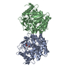


 Mass: 59.044 Da / Num. of mol.: 3 / Source method: obtained synthetically / Formula: C2H3O2
 Mass: 18.015 Da / Num. of mol.: 429 / Source method: isolated from a natural source / Formula: H2O
 Sample preparation
 Processing