 Movie
Movie Controller


+ Open data

- Basic information
| Entry | Database: PDB / ID: 3t0v | |||||||||
|---|---|---|---|---|---|---|---|---|---|---|
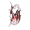
| Title | Unliganded fluorogen activating protein M8VL | |||||||||
 Components Components | immunoglobulin variable lambda domain | |||||||||
 Keywords Keywords | DYE-BINDING PROTEIN / immunoglobulin fold / fluorogen activation / dimethylindole red | |||||||||
| Function / homology | Immunoglobulins / Immunoglobulin-like / Sandwich / Mainly Beta / Chem-PE3 Function and homology information Function and homology information | |||||||||
| Biological species |  Homo sapiens (human) Homo sapiens (human) | |||||||||
| Method |  X-RAY DIFFRACTION / SYNCHROTRON / MOLECULAR REPLACEMENT / Resolution: 1.451 Å X-RAY DIFFRACTION / SYNCHROTRON / MOLECULAR REPLACEMENT / Resolution: 1.451 Å | |||||||||
 Authors Authors | Stanfield, R. / Senutovitch, N. / Bhattacharyya, S. / Rule, G. / Wilson, I.A. / Armitage, B. / Waggoner, A.S. / Berget, P. | |||||||||
 Citation Citation | Journal: Biochemistry / Year: 2012 Title: A variable light domain fluorogen activating protein homodimerizes to activate dimethylindole red. Authors: Senutovitch, N. / Stanfield, R.L. / Bhattacharyya, S. / Rule, G.S. / Wilson, I.A. / Armitage, B.A. / Waggoner, A.S. / Berget, P.B. | |||||||||
| History |
|
- Structure visualization
| Structure viewer | Molecule: MolmilJmol/JSmol |
|---|
- Downloads & links
-Download
| PDBx/mmCIF format | 3t0v.cif.gz | 63.7 KB | Display | PDBx/mmCIF format |
|---|---|---|---|---|
| PDB format | pdb3t0v.ent.gz | 44.9 KB | Display | PDB format |
| PDBx/mmJSON format | 3t0v.json.gz | Tree view | PDBx/mmJSON format | |
| Others |  Other downloads Other downloads |
-Validation report
| Arichive directory | https://data.pdbj.org/pub/pdb/validation_reports/t0/3t0vftp://data.pdbj.org/pub/pdb/validation_reports/t0/3t0v | HTTPS FTP |
|---|
-Related structure data


-Links
 PDBj
PDBj

- Assembly
| Deposited unit | 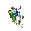
| ||||||||
|---|---|---|---|---|---|---|---|---|---|
| 1 |
| ||||||||
| Unit cell |
|
-Components
-Antibody , 1 types, 1 molecules A
| #1: Antibody | Mass: 13204.214 Da / Num. of mol.: 1 Source method: isolated from a genetically manipulated source Source: (gene. exp.) Homo sapiens (human) / Plasmid: pAK400 / Production host:  |
|---|
-Non-polymers , 6 types, 134 molecules 


| #2: Chemical | ChemComp-1PE /  Mass: 238.278 Da / Num. of mol.: 1 / Source method: obtained synthetically / Formula: C10H22O6 / Comment: precipitant*YM Mass: 238.278 Da / Num. of mol.: 1 / Source method: obtained synthetically / Formula: C10H22O6 / Comment: precipitant*YM | ||||||
|---|---|---|---|---|---|---|---|
| #3: Chemical | ChemComp-PE3 /  Mass: 634.751 Da / Num. of mol.: 1 / Source method: obtained synthetically / Formula: C28H58O15 Mass: 634.751 Da / Num. of mol.: 1 / Source method: obtained synthetically / Formula: C28H58O15 | ||||||
| #4: Chemical |  Mass: 62.068 Da / Num. of mol.: 3 / Source method: obtained synthetically / Formula: C2H6O2 Mass: 62.068 Da / Num. of mol.: 3 / Source method: obtained synthetically / Formula: C2H6O2#5: Chemical | ChemComp-SO4 / |  Mass: 96.063 Da / Num. of mol.: 1 / Source method: obtained synthetically / Formula: SO4 Mass: 96.063 Da / Num. of mol.: 1 / Source method: obtained synthetically / Formula: SO4#6: Chemical | ChemComp-TRS / |  Mass: 122.143 Da / Num. of mol.: 1 / Source method: obtained synthetically / Formula: C4H12NO3 / Comment: pH buffer*YM Mass: 122.143 Da / Num. of mol.: 1 / Source method: obtained synthetically / Formula: C4H12NO3 / Comment: pH buffer*YM#7: Water | ChemComp-HOH / | Mass: 18.015 Da / Num. of mol.: 127 / Source method: isolated from a natural source / Formula: H2O |
-Details
| Has protein modification | Y |
|---|
-Experimental details
-Experiment
| Experiment | Method: X-RAY DIFFRACTION / Number of used crystals: 1 |
|---|
- Sample preparation
| Crystal | Density Matthews: 2.27 Å3/Da / Density % sol: 45.75 % |
|---|---|
| Crystal grow | Temperature: 298 K / Method: vapor diffusion, sitting drop / pH: 7.5 Details: 2M ammonium sulfate, 2% PEG 400, 0.1M Hepes, pH 7.5, VAPOR DIFFUSION, SITTING DROP, temperature 298K |
-Data collection
| Diffraction | Mean temperature: 100 K |
|---|---|
| Diffraction source | Source: SYNCHROTRON / Site: ALS  / Beamline: 4.2.2 / Wavelength: 1 Å / Beamline: 4.2.2 / Wavelength: 1 Å |
| Detector | Type: NOIR-1 / Detector: CCD / Date: Aug 28, 2009 |
| Radiation | Protocol: SINGLE WAVELENGTH / Monochromatic (M) / Laue (L): M / Scattering type: x-ray |
| Radiation wavelength | Wavelength: 1 Å / Relative weight: 1 |
| Reflection | Resolution: 1.45→29.244 Å / Num. all: 21290 / Num. obs: 21290 / % possible obs: 97.7 % / Observed criterion σ(F): 0 / Observed criterion σ(I): -3 / Redundancy: 3.8 % / Biso Wilson estimate: 15.1 Å2 / Rsym value: 4.3 / Net I/σ(I): 30.1 |
| Reflection shell | Resolution: 1.45→1.48 Å / Redundancy: 2.7 % / Mean I/σ(I) obs: 2.4 / Num. unique all: 830 / Rsym value: 31.7 / % possible all: 79.2 |
- Processing
| Software |
| |||||||||||||||||||||||||||||||||||||||||||||||||||||||||||||||
|---|---|---|---|---|---|---|---|---|---|---|---|---|---|---|---|---|---|---|---|---|---|---|---|---|---|---|---|---|---|---|---|---|---|---|---|---|---|---|---|---|---|---|---|---|---|---|---|---|---|---|---|---|---|---|---|---|---|---|---|---|---|---|---|---|
| Refinement | Method to determine structure: MOLECULAR REPLACEMENT / Resolution: 1.451→29.2 Å / SU ML: 0.17 / σ(F): 0 / Phase error: 21.93 / Stereochemistry target values: ML
| |||||||||||||||||||||||||||||||||||||||||||||||||||||||||||||||
| Solvent computation | Shrinkage radii: 0.9 Å / VDW probe radii: 1.11 Å / Solvent model: FLAT BULK SOLVENT MODEL / Bsol: 52.32 Å2 / ksol: 0.379 e/Å3 | |||||||||||||||||||||||||||||||||||||||||||||||||||||||||||||||
| Displacement parameters |
| |||||||||||||||||||||||||||||||||||||||||||||||||||||||||||||||
| Refinement step | Cycle: LAST / Resolution: 1.451→29.2 Å
| |||||||||||||||||||||||||||||||||||||||||||||||||||||||||||||||
| Refine LS restraints |
| |||||||||||||||||||||||||||||||||||||||||||||||||||||||||||||||
| LS refinement shell |
|
