 Movie
Movie Controller

[English] 日本語
 Yorodumi
- PDB-4m3j: Structure of a single-domain camelid antibody fragment cAb-H7S sp... -
+ Open data

- Basic information
| Entry | Database: PDB / ID: 4m3j | ||||||
|---|---|---|---|---|---|---|---|
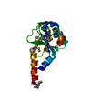
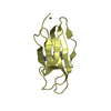
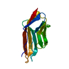
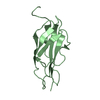
| Title | Structure of a single-domain camelid antibody fragment cAb-H7S specific of the BlaP beta-lactamase from Bacillus licheniformis | ||||||
 Components Components | Camelid heavy-chain antibody variable fragment cAb-H7S | ||||||
 Keywords Keywords | IMMUNE SYSTEM / Immunoglobulin fold / Antigen binding / beta-lactamase binding antibody | ||||||
| Function / homology | Immunoglobulins / Immunoglobulin-like / Sandwich / Mainly Beta Function and homology information Function and homology information | ||||||
| Biological species |  | ||||||
| Method |  X-RAY DIFFRACTION / SYNCHROTRON / MOLECULAR REPLACEMENT / Resolution: 1.95 Å X-RAY DIFFRACTION / SYNCHROTRON / MOLECULAR REPLACEMENT / Resolution: 1.95 Å | ||||||
 Authors Authors | Pain, C. / Kerff, F. / Herman, R. / Sauvage, E. / Preumont, S. / Charlier, P. / Dumoulin, M. | ||||||
 Citation Citation | Journal: To be Published Title: Probing the mechanism of aggregation of polyQ model proteins with camelid heavy-chain antibody fragments Authors: Pain, C. / Cosolo, A. / Preumont, S. / Scarafone, N. / Thorn, D. / Herman, R. / Spiegel, H. / Pardon, E. / Matagne, A. / Charlier, P. / Steyaert, J. / Damblon, C. / Kerff, F. / Esposito, G. / Dumoulin, M. | ||||||
| History |
|
- Structure visualization
| Structure viewer | Molecule: MolmilJmol/JSmol |
|---|
- Downloads & links
-Download
| PDBx/mmCIF format | 4m3j.cif.gz | 100.3 KB | Display | PDBx/mmCIF format |
|---|---|---|---|---|
| PDB format | pdb4m3j.ent.gz | 77.8 KB | Display | PDB format |
| PDBx/mmJSON format | 4m3j.json.gz | Tree view | PDBx/mmJSON format | |
| Others |  Other downloads Other downloads |
-Validation report
| Arichive directory | https://data.pdbj.org/pub/pdb/validation_reports/m3/4m3jftp://data.pdbj.org/pub/pdb/validation_reports/m3/4m3j | HTTPS FTP |
|---|
-Related structure data


-Links
 PDBj
PDBj

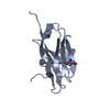
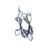
- Assembly
| Deposited unit | 
| ||||||||
|---|---|---|---|---|---|---|---|---|---|
| 1 | 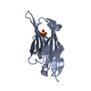
| ||||||||
| 2 | 
| ||||||||
| 3 |
| ||||||||
| Unit cell |
|
-Components
| #1: Antibody | Mass: 13524.937 Da / Num. of mol.: 2 Source method: isolated from a genetically manipulated source Source: (gene. exp.)  #2: Chemical |   Mass: 96.063 Da / Num. of mol.: 2 / Source method: obtained synthetically / Formula: SO4 Mass: 96.063 Da / Num. of mol.: 2 / Source method: obtained synthetically / Formula: SO4#3: Water | ChemComp-HOH / |  Mass: 18.015 Da / Num. of mol.: 42 / Source method: isolated from a natural source / Formula: H2O Mass: 18.015 Da / Num. of mol.: 42 / Source method: isolated from a natural source / Formula: H2OHas protein modification | Y | |
|---|
-Experimental details
-Experiment
| Experiment | Method: X-RAY DIFFRACTION / Number of used crystals: 1 |
|---|
- Sample preparation
| Crystal | Density Matthews: 2.26 Å3/Da / Density % sol: 45.48 % |
|---|---|
| Crystal grow | Temperature: 293 K / Method: vapor diffusion, sitting drop / pH: 4.6 Details: 0.2 uL 0.2 M ammonium sulphate 0.1 M NaAc, pH 4.6, 25% PEG4000 + 0.2 uL cAb-H7S 15 mg/ml 20 mM tris, pH 7.5, VAPOR DIFFUSION, SITTING DROP, temperature 293K |
-Data collection
| Diffraction | Mean temperature: 100 K |
|---|---|
| Diffraction source | Source: SYNCHROTRON / Site: SOLEIL  / Beamline: PROXIMA 1 / Wavelength: 0.98011 Å / Beamline: PROXIMA 1 / Wavelength: 0.98011 Å |
| Detector | Type: DECTRIS PILATUS 6M / Detector: PIXEL / Date: Feb 22, 2013 |
| Radiation | Protocol: SINGLE WAVELENGTH / Monochromatic (M) / Laue (L): M / Scattering type: x-ray |
| Radiation wavelength | Wavelength: 0.98011 Å / Relative weight: 1 |
| Reflection | Resolution: 1.95→45.7 Å / Num. all: 18390 / Num. obs: 18390 / % possible obs: 99.3 % / Redundancy: 6.9 % / Biso Wilson estimate: 48.7 Å2 / Rmerge(I) obs: 0.057 / Net I/σ(I): 16.2 |
| Reflection shell | Resolution: 1.95→2.05 Å / Redundancy: 6.8 % / Rmerge(I) obs: 0.818 / Mean I/σ(I) obs: 2.5 / Num. unique all: 2514 / % possible all: 95.5 |
- Processing
| Software |
| ||||||||||||||||||||||||||||||||||||||||||||||||||||||||||||||||||||||||||||||||||||||||||||||||||||||||||||||||||||||||||||||||||||||||||||||||||||||||||||||||||||||||||||||||||||||
|---|---|---|---|---|---|---|---|---|---|---|---|---|---|---|---|---|---|---|---|---|---|---|---|---|---|---|---|---|---|---|---|---|---|---|---|---|---|---|---|---|---|---|---|---|---|---|---|---|---|---|---|---|---|---|---|---|---|---|---|---|---|---|---|---|---|---|---|---|---|---|---|---|---|---|---|---|---|---|---|---|---|---|---|---|---|---|---|---|---|---|---|---|---|---|---|---|---|---|---|---|---|---|---|---|---|---|---|---|---|---|---|---|---|---|---|---|---|---|---|---|---|---|---|---|---|---|---|---|---|---|---|---|---|---|---|---|---|---|---|---|---|---|---|---|---|---|---|---|---|---|---|---|---|---|---|---|---|---|---|---|---|---|---|---|---|---|---|---|---|---|---|---|---|---|---|---|---|---|---|---|---|---|---|
| Refinement | Method to determine structure: MOLECULAR REPLACEMENT / Resolution: 1.95→45.7 Å / Cor.coef. Fo:Fc: 0.96 / Cor.coef. Fo:Fc free: 0.941 / SU B: 8.72 / SU ML: 0.119 / Cross valid method: THROUGHOUT / ESU R: 0.171 / ESU R Free: 0.161 / Stereochemistry target values: MAXIMUM LIKELIHOOD / Details: HYDROGENS HAVE BEEN USED IF PRESENT IN THE INPUT
| ||||||||||||||||||||||||||||||||||||||||||||||||||||||||||||||||||||||||||||||||||||||||||||||||||||||||||||||||||||||||||||||||||||||||||||||||||||||||||||||||||||||||||||||||||||||
| Solvent computation | Ion probe radii: 0.8 Å / Shrinkage radii: 0.8 Å / VDW probe radii: 1.2 Å / Solvent model: MASK | ||||||||||||||||||||||||||||||||||||||||||||||||||||||||||||||||||||||||||||||||||||||||||||||||||||||||||||||||||||||||||||||||||||||||||||||||||||||||||||||||||||||||||||||||||||||
| Displacement parameters | Biso mean: 44.937 Å2
| ||||||||||||||||||||||||||||||||||||||||||||||||||||||||||||||||||||||||||||||||||||||||||||||||||||||||||||||||||||||||||||||||||||||||||||||||||||||||||||||||||||||||||||||||||||||
| Refinement step | Cycle: LAST / Resolution: 1.95→45.7 Å
| ||||||||||||||||||||||||||||||||||||||||||||||||||||||||||||||||||||||||||||||||||||||||||||||||||||||||||||||||||||||||||||||||||||||||||||||||||||||||||||||||||||||||||||||||||||||
| Refine LS restraints |
|