[English] 日本語
Yorodumi
- PDB-3syj: Crystal structure of the Haemophilus influenzae Hap adhesin -

+
Open data


ID or keywords:

Loading...

-
Basic information

Entry
Database: PDB / ID: 3syj
TitleCrystal structure of the Haemophilus influenzae Hap adhesin
ComponentsAdhesion and penetration protein autotransporter
KeywordsCELL ADHESION / Bacterial aggregation and biofilm formation / self-associating autotransporter (SAAT) / oligomerization / beta helix fold / membrane
Function / homology
Function and homology information


Hydrolases; Acting on peptide bonds (peptidases); Serine endopeptidases / cell outer membrane / membrane => GO:0016020 / periplasmic space / cell adhesion / serine-type endopeptidase activity / cell surface / extracellular region / identical protein binding
Similarity search - Function
Pertactin, central region / Pertactin / Pectate Lyase C-like - #20 / Peptidase S6, IgA endopeptidase / Peptidase family S6 domain / Immunoglobulin A1 protease / Peptidase family S6 domain profile. / Outer membrane autotransporter barrel / Autotransporter beta-domain / Autotransporter beta-domain profile. ...Pertactin, central region / Pertactin / Pectate Lyase C-like - #20 / Peptidase S6, IgA endopeptidase / Peptidase family S6 domain / Immunoglobulin A1 protease / Peptidase family S6 domain profile. / Outer membrane autotransporter barrel / Autotransporter beta-domain / Autotransporter beta-domain profile. / Autotransporter beta-domain / Autotransporter beta-domain superfamily / Autotransporter, pectate lyase C-like domain superfamily / Thrombin, subunit H - #120 / Pectate Lyase C-like / Pectin lyase fold/virulence factor / 3 Solenoid / Serine proteases, trypsin family, serine active site / Thrombin, subunit H / Beta Barrel / Mainly Beta
Similarity search - Domain/homology
Adhesion and penetration protein autotransporter
Similarity search - Component
Biological speciesHaemophilus influenzae (bacteria)
MethodX-RAY DIFFRACTION / SYNCHROTRON / MOLECULAR REPLACEMENT / Resolution: 2.2 Å
AuthorsMeng, G.
CitationJournal: Embo J. / Year: 2011
Title: Crystal structure of the Haemophilus influenzae Hap adhesin reveals an intercellular oligomerization mechanism for bacterial aggregation
Authors: Meng, G. / Spahich, N. / Kenjale, R. / Waksman, G. / St Geme III, J.W.
History
DepositionJul 18, 2011Deposition site: RCSB / Processing site: PDBJ
Revision 1.0Aug 24, 2011Provider: repository / Type: Initial release
Revision 1.1Jul 3, 2013Group: Database references
Revision 1.2Nov 8, 2017Group: Refinement description / Category: software
Revision 1.3Nov 1, 2023Group: Data collection / Database references / Refinement description
Category: chem_comp_atom / chem_comp_bond ...chem_comp_atom / chem_comp_bond / database_2 / pdbx_initial_refinement_model
Item: _database_2.pdbx_DOI / _database_2.pdbx_database_accession

-
Structure visualization

Structure viewerMolecule:
MolmilJmol/JSmol

Downloads & links

-
Assembly

Deposited unit
A: Adhesion and penetration protein autotransporter


Theoretical massNumber of molelcules
Total (without water)112,1391
Polymers112,1391
Non-polymers00
Water14,106783
1
A: Adhesion and penetration protein autotransporter

A: Adhesion and penetration protein autotransporter


Theoretical massNumber of molelcules
Total (without water)224,2772
Polymers224,2772
Non-polymers00
Water362
TypeNameSymmetry operationNumber
identity operation1_555x,y,z1
crystal symmetry operation4_465x-1/2,-y+3/2,-z1
Unit cell
Length a, b, c (Å)41.410, 137.200, 209.580
Angle α, β, γ (deg.)90.00, 90.00, 90.00
Int Tables number19
Space group name H-MP212121
Details
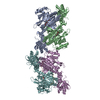
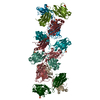
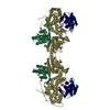
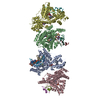
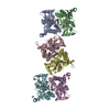
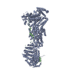
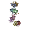
The second HapS of the biological assembly mediated by the C-terminal SAAT domain is generated alone the p21 a-axis. The crystal packing reveals a remarkable oligomerization of (HapS-HapS)n related by a crystallographic two-fold screw axis (i.e. p21 a-axis), perpendicular to the axis of the helix.

-
Components

#1: Protein Adhesion and penetration protein autotransporter / Hap adhesin / Adhesion and penetration protein / Adhesion and penetration protein translocator


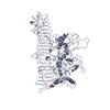
Mass: 112138.531 Da / Num. of mol.: 1 / Fragment: Hap passenger domain (HapS), UNP residues 26-1036
Source method: isolated from a genetically manipulated source
Source: (gene. exp.) Haemophilus influenzae (bacteria) / Strain: DB117 / Gene: hap / Plasmid: pJS106 / Production host: Escherichia coli (E. coli)
References: UniProt: P45387, Hydrolases; Acting on peptide bonds (peptidases); Serine endopeptidases
#2: Water ChemComp-HOH / water / Water


Mass: 18.015 Da / Num. of mol.: 783 / Source method: isolated from a natural source / Formula: H2O

-
Experimental details

-
Experiment

ExperimentMethod: X-RAY DIFFRACTION / Number of used crystals: 1

-
Sample preparation

CrystalDensity Matthews: 2.65 Å3/Da / Density % sol: 53.66 %
Crystal growTemperature: 298 K / Method: vapor diffusion, hanging drop / pH: 5.6
Details: 100mM sodium citrate, 14% (w/v) PEG 4000, 100mM ammonium sulfate, pH 5.6, VAPOR DIFFUSION, HANGING DROP, temperature 298K

-
Data collection

DiffractionMean temperature: 100 K
Diffraction sourceSource: SYNCHROTRON / Site: SSRF / Beamline: BL17U / Wavelength: 0.9763 Å
DetectorType: MARMOSAIC 225 mm CCD / Detector: CCD / Date: Apr 14, 2010
RadiationMonochromator: Graphite / Protocol: SINGLE WAVELENGTH / Monochromatic (M) / Laue (L): M / Scattering type: x-ray
Radiation wavelengthWavelength: 0.9763 Å / Relative weight: 1
ReflectionResolution: 2.2→68.6 Å / Num. obs: 60596 / % possible obs: 97.6 % / Observed criterion σ(F): 1 / Observed criterion σ(I): 1 / Redundancy: 4.8 % / Biso Wilson estimate: 31.8 Å2 / Rsym value: 0.135 / Net I/σ(I): 7.7
Reflection shellResolution: 2.2→2.32 Å / Redundancy: 2.8 % / Rsym value: 0.588 / % possible all: 89

-
Processing

Software
NameVersionClassification
DNAdata collection
PHASERphasing
PHENIX(phenix.refine)refinement
MOSFLMdata reduction
SCALAdata scaling
RefinementMethod to determine structure: MOLECULAR REPLACEMENT
Starting model: PDB ENTRIES 3H09 and 2WXR
Resolution: 2.2→68.6 Å / SU ML: 0.31 / σ(F): 0.01 / Phase error: 22.55 / Stereochemistry target values: Engh & Huber
RfactorNum. reflection% reflectionSelection details
Rfree0.2258 5174 5.1 %RANDOM
Rwork0.1865 ---
obs0.1885 60596 86.68 %-
all-60596 --
Solvent computationShrinkage radii: 0.9 Å / VDW probe radii: 1.11 Å / Solvent model: FLAT BULK SOLVENT MODEL / Bsol: 32.408 Å2 / ksol: 0.354 e/Å3
Displacement parameters
Baniso -1Baniso -2Baniso -3
1-1.8455 Å2-0 Å20 Å2
2---5.9874 Å2-0 Å2
3---4.1419 Å2
Refinement stepCycle: LAST / Resolution: 2.2→68.6 Å
ProteinNucleic acidLigandSolventTotal
Num. atoms7185 0 0 783 7968
Refine LS restraints
Refine-IDTypeDev idealNumber
X-RAY DIFFRACTIONf_bond_d0.0057337
X-RAY DIFFRACTIONf_angle_d0.9189928
X-RAY DIFFRACTIONf_dihedral_angle_d17.6872640
X-RAY DIFFRACTIONf_chiral_restr0.061073
X-RAY DIFFRACTIONf_plane_restr0.0031320
LS refinement shell
Resolution (Å)Rfactor RfreeNum. reflection RfreeRfactor RworkNum. reflection RworkRefine-ID% reflection obs (%)
2.2-2.2250.31351120.31311983X-RAY DIFFRACTION54
2.225-2.25120.34641150.33192154X-RAY DIFFRACTION59
2.2512-2.27860.39631590.32522272X-RAY DIFFRACTION61
2.2786-2.30750.3281580.29352446X-RAY DIFFRACTION66
2.3075-2.33790.32241220.27872537X-RAY DIFFRACTION69
2.3379-2.36990.30941240.28372677X-RAY DIFFRACTION73
2.3699-2.40370.29691390.26532930X-RAY DIFFRACTION78
2.4037-2.43960.321710.26653066X-RAY DIFFRACTION81
2.4396-2.47780.28391750.25273052X-RAY DIFFRACTION84
2.4778-2.51840.31511560.23633249X-RAY DIFFRACTION88
2.5184-2.56180.2861520.22843356X-RAY DIFFRACTION89
2.5618-2.60840.2561760.23353328X-RAY DIFFRACTION90
2.6084-2.65860.29241620.21613290X-RAY DIFFRACTION90
2.6586-2.71280.29151660.20643438X-RAY DIFFRACTION92
2.7128-2.77180.26261870.20223419X-RAY DIFFRACTION92
2.7718-2.83630.22171850.20263410X-RAY DIFFRACTION93
2.8363-2.90720.22482140.19963464X-RAY DIFFRACTION94
2.9072-2.98580.22471870.19153534X-RAY DIFFRACTION94
2.9858-3.07370.24132090.19183402X-RAY DIFFRACTION94
3.0737-3.17290.22822030.17733615X-RAY DIFFRACTION95
3.1729-3.28630.23471510.17143521X-RAY DIFFRACTION96
3.2863-3.41790.21731890.15153633X-RAY DIFFRACTION96
3.4179-3.57340.17392320.13563507X-RAY DIFFRACTION97
3.5734-3.76180.15772210.13463597X-RAY DIFFRACTION97
3.7618-3.99750.17691880.13063553X-RAY DIFFRACTION97
3.9975-4.30610.15891970.1243564X-RAY DIFFRACTION97
4.3061-4.73930.15151890.11893590X-RAY DIFFRACTION97
4.7393-5.42490.17371840.133558X-RAY DIFFRACTION97
5.4249-6.83380.17041780.16213600X-RAY DIFFRACTION95
6.8338-68.63330.21161730.18153551X-RAY DIFFRACTION96

+
About Yorodumi

-
News

-
Feb 9, 2022. New format data for meta-information of EMDB entries

New format data for meta-information of EMDB entries

  • Version 3 of the EMDB header file is now the official format.
  • The previous official version 1.9 will be removed from the archive.

Related info.:EMDB header

External links:wwPDB to switch to version 3 of the EMDB data model

-
Aug 12, 2020. Covid-19 info

Covid-19 info

URL: https://pdbj.org/emnavi/covid19.php

New page: Covid-19 featured information page in EM Navigator.

Related info.:Covid-19 info / Mar 5, 2020. Novel coronavirus structure data

+
Mar 5, 2020. Novel coronavirus structure data

Novel coronavirus structure data

Related info.:Yorodumi Speices / Aug 12, 2020. Covid-19 info

External links:COVID-19 featured content - PDBj / Molecule of the Month (242):Coronavirus Proteases

+
Jan 31, 2019. EMDB accession codes are about to change! (news from PDBe EMDB page)

EMDB accession codes are about to change! (news from PDBe EMDB page)

  • The allocation of 4 digits for EMDB accession codes will soon come to an end. Whilst these codes will remain in use, new EMDB accession codes will include an additional digit and will expand incrementally as the available range of codes is exhausted. The current 4-digit format prefixed with “EMD-” (i.e. EMD-XXXX) will advance to a 5-digit format (i.e. EMD-XXXXX), and so on. It is currently estimated that the 4-digit codes will be depleted around Spring 2019, at which point the 5-digit format will come into force.
  • The EM Navigator/Yorodumi systems omit the EMD- prefix.

Related info.:Q: What is EMD? / ID/Accession-code notation in Yorodumi/EM Navigator

External links:EMDB Accession Codes are Changing Soon! / Contact to PDBj

+
Jul 12, 2017. Major update of PDB

Major update of PDB

  • wwPDB released updated PDB data conforming to the new PDBx/mmCIF dictionary.
  • This is a major update changing the version number from 4 to 5, and with Remediation, in which all the entries are updated.
  • In this update, many items about electron microscopy experimental information are reorganized (e.g. em_software).
  • Now, EM Navigator and Yorodumi are based on the updated data.

External links:wwPDB Remediation / Enriched Model Files Conforming to OneDep Data Standards Now Available in the PDB FTP Archive

-
Yorodumi

Thousand views of thousand structures

  • Yorodumi is a browser for structure data from EMDB, PDB, SASBDB, etc.
  • This page is also the successor to EM Navigator detail page, and also detail information page/front-end page for Omokage search.
  • The word "yorodu" (or yorozu) is an old Japanese word meaning "ten thousand". "mi" (miru) is to see.

Related info.:EMDB / PDB / SASBDB / Comparison of 3 databanks / Yorodumi Search / Aug 31, 2016. New EM Navigator & Yorodumi / Yorodumi Papers / Jmol/JSmol / Function and homology information / Changes in new EM Navigator and Yorodumi

Read more