 Movie
Movie Controller

[English] 日本語
 Yorodumi
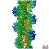
- PDB-5u1t: Crystal structure of the Saccharomyces cerevisiae separase-securi... -
+ Open data

- Basic information
| Entry | Database: PDB / ID: 5u1t | |||||||||
|---|---|---|---|---|---|---|---|---|---|---|
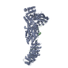
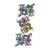
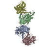
| Title | Crystal structure of the Saccharomyces cerevisiae separase-securin complex at 2.6 angstrom resolution | |||||||||
 Components Components |
| |||||||||
 Keywords Keywords | HYDROLASE / separase-securin complex / ESP1 / PDS1 / chromosome segregation / cohesion / helical region / inhibition / chaperon | |||||||||
| Function / homology |  Function and homology information Function and homology informationmeiosis II / positive regulation of metaphase/anaphase transition of meiotic cell cycle / separase-securin complex / Separation of Sister Chromatids / negative regulation of exit from mitosis / separase / negative regulation of protein localization to nucleolus / regulation of mitotic spindle elongation / meiotic chromosome separation / mitotic spindle pole body ...meiosis II / positive regulation of metaphase/anaphase transition of meiotic cell cycle / separase-securin complex / Separation of Sister Chromatids / negative regulation of exit from mitosis / separase / negative regulation of protein localization to nucleolus / regulation of mitotic spindle elongation / meiotic chromosome separation / mitotic spindle pole body / positive regulation of exit from mitosis / meiosis I / recombinational repair / mitotic sister chromatid segregation / spindle / mitotic spindle / intracellular protein localization / cell division / cysteine-type endopeptidase activity / apoptotic process / DNA damage response / enzyme binding / mitochondrion / proteolysis / nucleus / cytoplasm Similarity search - Function | |||||||||
| Biological species |  | |||||||||
| Method |  X-RAY DIFFRACTION / SYNCHROTRON / MOLECULAR REPLACEMENT / Resolution: 2.6 Å X-RAY DIFFRACTION / SYNCHROTRON / MOLECULAR REPLACEMENT / Resolution: 2.6 Å | |||||||||
 Authors Authors | Luo, S. / Tong, L. | |||||||||
| Funding support |  United States, 2items United States, 2items
| |||||||||
 Citation Citation | Journal: Nature / Year: 2017 Title: Molecular mechanism for the regulation of yeast separase by securin. Authors: Luo, S. / Tong, L. | |||||||||
| History |
|
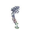
- Structure visualization
| Structure viewer | Molecule: MolmilJmol/JSmol |
|---|
- Downloads & links
-Download
| PDBx/mmCIF format | 5u1t.cif.gz | 329 KB | Display | PDBx/mmCIF format |
|---|---|---|---|---|
| PDB format | pdb5u1t.ent.gz | 257.1 KB | Display | PDB format |
| PDBx/mmJSON format | 5u1t.json.gz | Tree view | PDBx/mmJSON format | |
| Others |  Other downloads Other downloads |
-Validation report
| Arichive directory | https://data.pdbj.org/pub/pdb/validation_reports/u1/5u1tftp://data.pdbj.org/pub/pdb/validation_reports/u1/5u1t | HTTPS FTP |
|---|
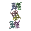
-Related structure data
| Related structure data |  5u1sSC S: Starting model for refinement C: citing same article ( |
|---|---|
| Similar structure data |
-Links
 PDBj
PDBj
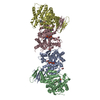
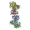
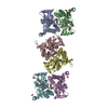
- Assembly
| Deposited unit | 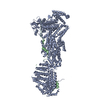
| ||||||||
|---|---|---|---|---|---|---|---|---|---|
| 1 |
| ||||||||
| Unit cell |
|
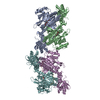
-Components
| #1: Protein | Mass: 184044.469 Da / Num. of mol.: 1 Fragment: UNP residues 71-1630, helical domain and catalytic domain Source method: isolated from a genetically manipulated source Source: (gene. exp.) Strain: ATCC 204508 / S288c / Gene: ESP1, YGR098C / Plasmid: pFL / Production host:  Trichoplusia ni (cabbage looper) / Strain (production host): High Five Cell / References: UniProt: Q03018, separase Trichoplusia ni (cabbage looper) / Strain (production host): High Five Cell / References: UniProt: Q03018, separase |
|---|---|
| #2: Protein | Mass: 13366.599 Da / Num. of mol.: 1 Fragment: UNP residues 258-373, separase interaction segment Source method: isolated from a genetically manipulated source Source: (gene. exp.) Strain: ATCC 204508 / S288c / Gene: PDS1, YDR113C, YD9727.08C / Plasmid: pFL / Production host: Trichoplusia ni (cabbage looper) / Strain (production host): High Five Cell / References: UniProt: P40316 |
| #3: Water | ChemComp-HOH /  Mass: 18.015 Da / Num. of mol.: 16 / Source method: isolated from a natural source / Formula: H2O Mass: 18.015 Da / Num. of mol.: 16 / Source method: isolated from a natural source / Formula: H2O |
| Has protein modification | Y |
-Experimental details
-Experiment
| Experiment | Method: X-RAY DIFFRACTION / Number of used crystals: 1 |
|---|
- Sample preparation
| Crystal | Density Matthews: 3.15 Å3/Da / Density % sol: 60.95 % |
|---|---|
| Crystal grow | Temperature: 293 K / Method: vapor diffusion / pH: 6 Details: 0.25 M Na/K-phosphate (pH 6.0) and 14% (w/v) PEG 3350 |
-Data collection
| Diffraction | Mean temperature: 100 K | ||||||||||||||||||||||||||||||||||||||||||||||||||||||||||||||||||
|---|---|---|---|---|---|---|---|---|---|---|---|---|---|---|---|---|---|---|---|---|---|---|---|---|---|---|---|---|---|---|---|---|---|---|---|---|---|---|---|---|---|---|---|---|---|---|---|---|---|---|---|---|---|---|---|---|---|---|---|---|---|---|---|---|---|---|---|
| Diffraction source | Source: SYNCHROTRON / Site: APS / Beamline: 24-ID-C / Wavelength: 0.9792 Å | ||||||||||||||||||||||||||||||||||||||||||||||||||||||||||||||||||
| Detector | Type: DECTRIS PILATUS 6M-F / Detector: PIXEL / Date: Aug 18, 2016 / Details: DECTRIS PILATUS 6M-F | ||||||||||||||||||||||||||||||||||||||||||||||||||||||||||||||||||
| Radiation | Protocol: SINGLE WAVELENGTH / Monochromatic (M) / Laue (L): M / Scattering type: x-ray | ||||||||||||||||||||||||||||||||||||||||||||||||||||||||||||||||||
| Radiation wavelength | Wavelength: 0.9792 Å / Relative weight: 1 | ||||||||||||||||||||||||||||||||||||||||||||||||||||||||||||||||||
| Reflection | Resolution: 2.6→50 Å / Num. obs: 77491 / % possible obs: 100 % / Redundancy: 10 % / Biso Wilson estimate: 62.34 Å2 / Rmerge(I) obs: 0.145 / Rpim(I) all: 0.059 / Rrim(I) all: 0.153 / Χ2: 1.06 / Net I/av σ(I): 14.42 / Net I/σ(I): 4.3 / Num. measured all: 774372 | ||||||||||||||||||||||||||||||||||||||||||||||||||||||||||||||||||
| Reflection shell |
|
- Processing
| Software |
| |||||||||||||||||||||||||||||||||||||||||||||||||||||||||||||||||||||||||||||||||||||||||||||||||||||||||
|---|---|---|---|---|---|---|---|---|---|---|---|---|---|---|---|---|---|---|---|---|---|---|---|---|---|---|---|---|---|---|---|---|---|---|---|---|---|---|---|---|---|---|---|---|---|---|---|---|---|---|---|---|---|---|---|---|---|---|---|---|---|---|---|---|---|---|---|---|---|---|---|---|---|---|---|---|---|---|---|---|---|---|---|---|---|---|---|---|---|---|---|---|---|---|---|---|---|---|---|---|---|---|---|---|---|---|
| Refinement | Method to determine structure: MOLECULAR REPLACEMENT Starting model: 5U1S Resolution: 2.6→48.667 Å / SU ML: 0.4 / Cross valid method: FREE R-VALUE / σ(F): 1.33 / Phase error: 32.01
| |||||||||||||||||||||||||||||||||||||||||||||||||||||||||||||||||||||||||||||||||||||||||||||||||||||||||
| Solvent computation | Shrinkage radii: 0.9 Å / VDW probe radii: 1.11 Å | |||||||||||||||||||||||||||||||||||||||||||||||||||||||||||||||||||||||||||||||||||||||||||||||||||||||||
| Displacement parameters | Biso max: 142.36 Å2 / Biso mean: 72.031 Å2 / Biso min: 35.38 Å2 | |||||||||||||||||||||||||||||||||||||||||||||||||||||||||||||||||||||||||||||||||||||||||||||||||||||||||
| Refinement step | Cycle: final / Resolution: 2.6→48.667 Å
| |||||||||||||||||||||||||||||||||||||||||||||||||||||||||||||||||||||||||||||||||||||||||||||||||||||||||
| Refine LS restraints |
| |||||||||||||||||||||||||||||||||||||||||||||||||||||||||||||||||||||||||||||||||||||||||||||||||||||||||
| LS refinement shell | Refine-ID: X-RAY DIFFRACTION / Total num. of bins used: 14
|
