 Movie
Movie Controller

[English] 日本語
 Yorodumi
- PDB-6ajb: Crystal structure of Trypanosoma brucei glycosomal isocitrate deh... -
+ Open data

- Basic information
| Entry | Database: PDB / ID: 6ajb | ||||||
|---|---|---|---|---|---|---|---|
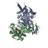
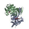
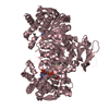
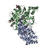
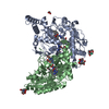
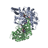
| Title | Crystal structure of Trypanosoma brucei glycosomal isocitrate dehydrogenase in complex with NADH, alpha-ketoglutarate and ca2+ | ||||||
 Components Components | Isocitrate dehydrogenase [NADP] | ||||||
 Keywords Keywords | OXIDOREDUCTASE / Trypanosoma brucei / glycosome / metabolism / isocitrate dehydrogenase | ||||||
| Function / homology |  Function and homology information Function and homology informationisocitrate metabolic process / isocitrate dehydrogenase (NADP+) / isocitrate dehydrogenase (NADP+) activity / NADP+ metabolic process / glycosome / tricarboxylic acid cycle / NAD binding / magnesium ion binding / mitochondrion / cytoplasm Similarity search - Function | ||||||
| Biological species |  | ||||||
| Method |  X-RAY DIFFRACTION / SYNCHROTRON / MOLECULAR REPLACEMENT / Resolution: 2.9 Å X-RAY DIFFRACTION / SYNCHROTRON / MOLECULAR REPLACEMENT / Resolution: 2.9 Å | ||||||
 Authors Authors | Wang, X. / Inaoka, D.K. / Shiba, T. / Balogun, E.O. / Ziebart, N. / Allman, S. / Watanabe, Y. / Nozaki, T. / Boshart, M. / Bringaud, F. ...Wang, X. / Inaoka, D.K. / Shiba, T. / Balogun, E.O. / Ziebart, N. / Allman, S. / Watanabe, Y. / Nozaki, T. / Boshart, M. / Bringaud, F. / Harada, S. / Kita, K. | ||||||
 Citation Citation | Journal: To Be Published Title: Biochemical characterization of a novel Trypanosoma brucei glycosomal isocitrate dehydrogenase with dual coenzyme specificity (NADP+/NAD+) Authors: Wang, X. / Inaoka, D.K. / Shiba, T. / Balogun, E.O. / Ziebart, N. / Allman, S. / Watanabe, Y. / Nozaki, T. / Boshart, M. / Bringaud, F. / Harada, S. / Kita, K. | ||||||
| History |
|
- Structure visualization
| Structure viewer | Molecule: MolmilJmol/JSmol |
|---|
- Downloads & links
-Download
| PDBx/mmCIF format | 6ajb.cif.gz | 649.1 KB | Display | PDBx/mmCIF format |
|---|---|---|---|---|
| PDB format | pdb6ajb.ent.gz | 544.1 KB | Display | PDB format |
| PDBx/mmJSON format | 6ajb.json.gz | Tree view | PDBx/mmJSON format | |
| Others |  Other downloads Other downloads |
-Validation report
| Arichive directory | https://data.pdbj.org/pub/pdb/validation_reports/aj/6ajbftp://data.pdbj.org/pub/pdb/validation_reports/aj/6ajb | HTTPS FTP |
|---|
-Related structure data
| Related structure data |  6aj6SC 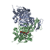 6aj8C 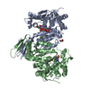 6ajaC  6ajcC S: Starting model for refinement C: citing same article ( |
|---|---|
| Similar structure data |


-Links
 PDBj
PDBj


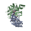
- Assembly
| Deposited unit | 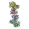
| ||||||||
|---|---|---|---|---|---|---|---|---|---|
| 1 | 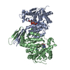
| ||||||||
| 2 | 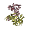
| ||||||||
| Unit cell |
|
-Components
| #1: Protein | Mass: 46791.590 Da / Num. of mol.: 4 / Mutation: M1S Source method: isolated from a genetically manipulated source Source: (gene. exp.)  References: UniProt: Q387G0, isocitrate dehydrogenase (NADP+) #2: Chemical | ChemComp-NAD /   Mass: 663.425 Da / Num. of mol.: 4 / Source method: obtained synthetically / Formula: C21H27N7O14P2 / Comment: NAD*YM Mass: 663.425 Da / Num. of mol.: 4 / Source method: obtained synthetically / Formula: C21H27N7O14P2 / Comment: NAD*YM#3: Chemical | ChemComp-CA /   Mass: 40.078 Da / Num. of mol.: 4 / Source method: obtained synthetically / Formula: Ca Mass: 40.078 Da / Num. of mol.: 4 / Source method: obtained synthetically / Formula: Ca#4: Chemical | ChemComp-AKG / |   Mass: 146.098 Da / Num. of mol.: 1 / Source method: obtained synthetically / Formula: C5H6O5 Mass: 146.098 Da / Num. of mol.: 1 / Source method: obtained synthetically / Formula: C5H6O5Sequence details | The sequence was derived from Trypanosome brucei brucei ILtat 1.4. However, the sequence reference ...The sequence was derived from Trypanosome brucei brucei ILtat 1.4. However, the sequence reference available at uniprot at the time of data processing was derived from a different strain TRYPANOSOM | |
|---|
-Experimental details
-Experiment
| Experiment | Method: X-RAY DIFFRACTION / Number of used crystals: 1 |
|---|
- Sample preparation
| Crystal | Density Matthews: 2.54 Å3/Da / Density % sol: 51.54 % |
|---|---|
| Crystal grow | Temperature: 293 K / Method: vapor diffusion, sitting drop / pH: 7 / Details: 20% PEG 6000, 100 mM HEPES/NaOH pH 7.0, 200mM NaCl |
-Data collection
| Diffraction | Mean temperature: 100 K |
|---|---|
| Diffraction source | Source: SYNCHROTRON / Site: SPring-8  / Beamline: BL44XU / Wavelength: 0.9 Å / Beamline: BL44XU / Wavelength: 0.9 Å |
| Detector | Type: RAYONIX MX300HE / Detector: CCD / Date: Jun 16, 2016 |
| Radiation | Protocol: SINGLE WAVELENGTH / Monochromatic (M) / Laue (L): M / Scattering type: x-ray |
| Radiation wavelength | Wavelength: 0.9 Å / Relative weight: 1 |
| Reflection | Resolution: 2.9→50 Å / Num. obs: 39505 / % possible obs: 93.8 % / Redundancy: 2.4 % / Rmerge(I) obs: 0.098 / Net I/σ(I): 5.7 |
| Reflection shell | Resolution: 2.9→2.95 Å / Redundancy: 2.3 % / Rmerge(I) obs: 0.692 / Mean I/σ(I) obs: 1.03 / Num. unique obs: 1842 / % possible all: 89.2 |
- Processing
| Software |
| ||||||||||||||||||||||||||||||||||||||||||||||||||||||||||||||||||||||||||||||||||||||||||||||||||||||||||||||||||||||||||||||||||||||||||||||||||||||||||||||||||||||||||||||||||||||
|---|---|---|---|---|---|---|---|---|---|---|---|---|---|---|---|---|---|---|---|---|---|---|---|---|---|---|---|---|---|---|---|---|---|---|---|---|---|---|---|---|---|---|---|---|---|---|---|---|---|---|---|---|---|---|---|---|---|---|---|---|---|---|---|---|---|---|---|---|---|---|---|---|---|---|---|---|---|---|---|---|---|---|---|---|---|---|---|---|---|---|---|---|---|---|---|---|---|---|---|---|---|---|---|---|---|---|---|---|---|---|---|---|---|---|---|---|---|---|---|---|---|---|---|---|---|---|---|---|---|---|---|---|---|---|---|---|---|---|---|---|---|---|---|---|---|---|---|---|---|---|---|---|---|---|---|---|---|---|---|---|---|---|---|---|---|---|---|---|---|---|---|---|---|---|---|---|---|---|---|---|---|---|---|
| Refinement | Method to determine structure: MOLECULAR REPLACEMENT Starting model: 6AJ6 Resolution: 2.9→19.98 Å / Cor.coef. Fo:Fc: 0.936 / Cor.coef. Fo:Fc free: 0.839 / SU B: 57.731 / SU ML: 0.457 / Cross valid method: THROUGHOUT / ESU R Free: 0.569 / Details: HYDROGENS HAVE BEEN ADDED IN THE RIDING POSITIONS
| ||||||||||||||||||||||||||||||||||||||||||||||||||||||||||||||||||||||||||||||||||||||||||||||||||||||||||||||||||||||||||||||||||||||||||||||||||||||||||||||||||||||||||||||||||||||
| Solvent computation | Ion probe radii: 0.8 Å / Shrinkage radii: 0.8 Å / VDW probe radii: 1.2 Å | ||||||||||||||||||||||||||||||||||||||||||||||||||||||||||||||||||||||||||||||||||||||||||||||||||||||||||||||||||||||||||||||||||||||||||||||||||||||||||||||||||||||||||||||||||||||
| Displacement parameters | Biso mean: 56.642 Å2
| ||||||||||||||||||||||||||||||||||||||||||||||||||||||||||||||||||||||||||||||||||||||||||||||||||||||||||||||||||||||||||||||||||||||||||||||||||||||||||||||||||||||||||||||||||||||
| Refinement step | Cycle: 1 / Resolution: 2.9→19.98 Å
| ||||||||||||||||||||||||||||||||||||||||||||||||||||||||||||||||||||||||||||||||||||||||||||||||||||||||||||||||||||||||||||||||||||||||||||||||||||||||||||||||||||||||||||||||||||||
| Refine LS restraints |
|