 Movie
Movie Controller


+ Open data

- Basic information
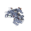
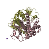
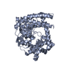
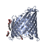
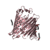
| Entry | Database: PDB / ID: 3syb | ||||||
|---|---|---|---|---|---|---|---|
| Title | Crystal structure of Pseudomonas aeruginosa OccD3 (OpdP) | ||||||
 Components Components | Glycine-glutamate dipeptide porin OpdP | ||||||
 Keywords Keywords | MEMBRANE PROTEIN / beta-barrel / channel / bacterial outer membrane | ||||||
| Function / homology |  Function and homology information Function and homology information | ||||||
| Biological species |   Pseudomonas aeruginosa (bacteria) Pseudomonas aeruginosa (bacteria) | ||||||
| Method |  X-RAY DIFFRACTION / SYNCHROTRON / P / Resolution: 2.7 Å X-RAY DIFFRACTION / SYNCHROTRON / P / Resolution: 2.7 Å | ||||||
 Authors Authors | van den Berg, B. / Eren, E. | ||||||
 Citation Citation | Journal: Plos Biol. / Year: 2012 Title: Substrate Specificity within a Family of Outer Membrane Carboxylate Channels. Authors: Eren, E. / Vijayaraghavan, J. / Liu, J. / Cheneke, B.R. / Touw, D.S. / Lepore, B.W. / Indic, M. / Movileanu, L. / van den Berg, B. | ||||||
| History |
|
- Structure visualization
| Structure viewer | Molecule: MolmilJmol/JSmol |
|---|
- Downloads & links
-Download
| PDBx/mmCIF format | 3syb.cif.gz | 93.5 KB | Display | PDBx/mmCIF format |
|---|---|---|---|---|
| PDB format | pdb3syb.ent.gz | 71 KB | Display | PDB format |
| PDBx/mmJSON format | 3syb.json.gz | Tree view | PDBx/mmJSON format | |
| Others |  Other downloads Other downloads |
-Validation report
| Arichive directory | https://data.pdbj.org/pub/pdb/validation_reports/sy/3sybftp://data.pdbj.org/pub/pdb/validation_reports/sy/3syb | HTTPS FTP |
|---|
-Related structure data
| Related structure data |  3sy7C  3sy9C 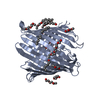 3sysC  3szdC  3szvC  3t0sC  3t20C 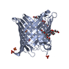 3t24C C: citing same article ( |
|---|---|
| Similar structure data |


-Links
 PDBj
PDBj- Assembly
| Deposited unit | 
| ||||||||
|---|---|---|---|---|---|---|---|---|---|
| 1 |
| ||||||||
| 2 | 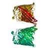
| ||||||||
| Unit cell |
| ||||||||
| Components on special symmetry positions |
|
-Components
| #1: Protein | Mass: 51457.297 Da / Num. of mol.: 1 / Fragment: unp residues 26-484 Source method: isolated from a genetically manipulated source Source: (gene. exp.) Pseudomonas aeruginosa (bacteria) / Gene: opdP, PA4501 / Production host: | ||||
|---|---|---|---|---|---|
| #2: Chemical |   Mass: 96.063 Da / Num. of mol.: 2 / Source method: obtained synthetically / Formula: SO4 Mass: 96.063 Da / Num. of mol.: 2 / Source method: obtained synthetically / Formula: SO4#3: Chemical |   Mass: 306.438 Da / Num. of mol.: 3 / Source method: obtained synthetically / Formula: C16H34O5 / Comment: C8E, detergent*YM Mass: 306.438 Da / Num. of mol.: 3 / Source method: obtained synthetically / Formula: C16H34O5 / Comment: C8E, detergent*YM#4: Water | ChemComp-HOH / |  Mass: 18.015 Da / Num. of mol.: 45 / Source method: isolated from a natural source / Formula: H2O Mass: 18.015 Da / Num. of mol.: 45 / Source method: isolated from a natural source / Formula: H2O |
-Experimental details
-Experiment
| Experiment | Method: X-RAY DIFFRACTION / Number of used crystals: 1 |
|---|
- Sample preparation
| Crystal | Density Matthews: 3.18 Å3/Da / Density % sol: 61.37 % |
|---|---|
| Crystal grow | Temperature: 295 K / Method: vapor diffusion, sitting drop / pH: 4 Details: 25% PEG 1000 0.2 M ammonium sulphate 50 mM Na-citrate, pH 4.0, VAPOR DIFFUSION, SITTING DROP, temperature 295K |
-Data collection
| Diffraction | Mean temperature: 100 K |
|---|---|
| Diffraction source | Source: SYNCHROTRON / Site: NSLS  / Beamline: X6A / Wavelength: 0.972 Å / Beamline: X6A / Wavelength: 0.972 Å |
| Detector | Type: ADSC QUANTUM 270 / Detector: CCD / Date: Feb 17, 2011 |
| Radiation | Monochromator: Si(111) / Protocol: SINGLE WAVELENGTH / Monochromatic (M) / Laue (L): M / Scattering type: x-ray |
| Radiation wavelength | Wavelength: 0.972 Å / Relative weight: 1 |
| Reflection | Resolution: 2.66→50 Å / Num. obs: 18213 / % possible obs: 92.9 % / Observed criterion σ(F): 0 / Observed criterion σ(I): -3 |
| Reflection shell | Resolution: 2.66→2.71 Å / % possible all: 96.5 |
- Processing
| Software |
| ||||||||||||||||||||||||||||||||||||||||||||||||||||||||||||||||||||||||||||||||||||||||||||||||||
|---|---|---|---|---|---|---|---|---|---|---|---|---|---|---|---|---|---|---|---|---|---|---|---|---|---|---|---|---|---|---|---|---|---|---|---|---|---|---|---|---|---|---|---|---|---|---|---|---|---|---|---|---|---|---|---|---|---|---|---|---|---|---|---|---|---|---|---|---|---|---|---|---|---|---|---|---|---|---|---|---|---|---|---|---|---|---|---|---|---|---|---|---|---|---|---|---|---|---|---|
| Refinement | Method to determine structure: P / Resolution: 2.7→9.993 Å / SU ML: 0.76 / σ(F): 1.34 / Phase error: 27.08 / Stereochemistry target values: ML
| ||||||||||||||||||||||||||||||||||||||||||||||||||||||||||||||||||||||||||||||||||||||||||||||||||
| Solvent computation | Shrinkage radii: 0.49 Å / VDW probe radii: 0.8 Å / Solvent model: FLAT BULK SOLVENT MODEL / Bsol: 43.632 Å2 / ksol: 0.368 e/Å3 | ||||||||||||||||||||||||||||||||||||||||||||||||||||||||||||||||||||||||||||||||||||||||||||||||||
| Displacement parameters |
| ||||||||||||||||||||||||||||||||||||||||||||||||||||||||||||||||||||||||||||||||||||||||||||||||||
| Refinement step | Cycle: LAST / Resolution: 2.7→9.993 Å
| ||||||||||||||||||||||||||||||||||||||||||||||||||||||||||||||||||||||||||||||||||||||||||||||||||
| Refine LS restraints |
| ||||||||||||||||||||||||||||||||||||||||||||||||||||||||||||||||||||||||||||||||||||||||||||||||||
| LS refinement shell |
|
