 Movie
Movie Controller


+ Open data

- Basic information
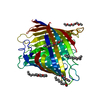
| Entry | Database: PDB / ID: 2y0l | ||||||
|---|---|---|---|---|---|---|---|
| Title | Crystal Structure of Pseudomonas Aeruginosa OpdO | ||||||
 Components Components | CIS-ACONITATE PORIN OPDH | ||||||
 Keywords Keywords | TRANSPORT PROTEIN / OUTER MEMBRANE / OPRD | ||||||
| Function / homology |  Function and homology information Function and homology information | ||||||
| Biological species |   PSEUDOMONAS AERUGINOSA (bacteria) PSEUDOMONAS AERUGINOSA (bacteria) | ||||||
| Method |  X-RAY DIFFRACTION / SYNCHROTRON / MOLECULAR REPLACEMENT / Resolution: 2.59 Å X-RAY DIFFRACTION / SYNCHROTRON / MOLECULAR REPLACEMENT / Resolution: 2.59 Å | ||||||
 Authors Authors | Touw, D.S. / Vijayaraghavan, J. / Vandenberg, B. | ||||||
 Citation Citation | Journal: To be Published Title: Crystal Structure of Pseudomonas Aeruginosa Opdo Authors: Touw, D.S. / Vijayaraghavan, J. / Vandenberg, B. | ||||||
| History |
| ||||||
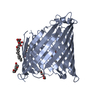
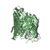
| Remark 700 | SHEET DETERMINATION METHOD: DSSP THE SHEETS PRESENTED AS "AA" IN EACH CHAIN ON SHEET RECORDS BELOW ... SHEET DETERMINATION METHOD: DSSP THE SHEETS PRESENTED AS "AA" IN EACH CHAIN ON SHEET RECORDS BELOW IS ACTUALLY AN 18-STRANDED BARREL THIS IS REPRESENTED BY A 19-STRANDED SHEET IN WHICH THE FIRST AND LAST STRANDS ARE IDENTICAL. |
- Structure visualization
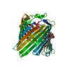
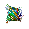
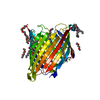
| Structure viewer | Molecule: MolmilJmol/JSmol |
|---|
- Downloads & links
-Download
| PDBx/mmCIF format | 2y0l.cif.gz | 85.7 KB | Display | PDBx/mmCIF format |
|---|---|---|---|---|
| PDB format | pdb2y0l.ent.gz | 64 KB | Display | PDB format |
| PDBx/mmJSON format | 2y0l.json.gz | Tree view | PDBx/mmJSON format | |
| Others |  Other downloads Other downloads |
-Validation report
| Arichive directory | https://data.pdbj.org/pub/pdb/validation_reports/y0/2y0lftp://data.pdbj.org/pub/pdb/validation_reports/y0/2y0l | HTTPS FTP |
|---|
-Related structure data
| Related structure data |  2y0kC  2qtkS S: Starting model for refinement C: citing same article ( |
|---|---|
| Similar structure data |


-Links
 PDBj
PDBj- Assembly
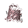
| Deposited unit | 
| ||||||||
|---|---|---|---|---|---|---|---|---|---|
| 1 |
| ||||||||
| Unit cell |
|
-Components
| #1: Protein | Mass: 44433.492 Da / Num. of mol.: 1 / Fragment: MATURE PROTEIN, RESIDUES 26-427 Source method: isolated from a genetically manipulated source Source: (gene. exp.) PSEUDOMONAS AERUGINOSA (bacteria) / Strain: PAO1 / Plasmid: PB22 / Production host: |
|---|---|
| #2: Water | ChemComp-HOH /  Mass: 18.015 Da / Num. of mol.: 97 / Source method: isolated from a natural source / Formula: H2O Mass: 18.015 Da / Num. of mol.: 97 / Source method: isolated from a natural source / Formula: H2O |
| Sequence details | AUTHORS INDICATE THAT THIS IS THE MATURE PROTEIN LACKING 25 RESIDUE SIGNAL PEPTIDE. |
-Experimental details
-Experiment
| Experiment | Method: X-RAY DIFFRACTION |
|---|
- Sample preparation
| Crystal | Density Matthews: 3.4 Å3/Da / Density % sol: 64 % / Description: NONE |
|---|---|
| Crystal grow | Details: 0.1 M LITHIUM SULFATE, 0.05 M DISODIUM PHOSPHATE PH 4, 33% PEG 400. |
-Data collection
| Diffraction | Mean temperature: 100 K |
|---|---|
| Diffraction source | Source: SYNCHROTRON / Site: NSLS  / Beamline: X6A / Wavelength: 1 / Beamline: X6A / Wavelength: 1 |
| Detector | Type: ADSC QUANTUM 210 / Detector: CCD / Date: Sep 15, 2010 |
| Radiation | Protocol: SINGLE WAVELENGTH / Monochromatic (M) / Laue (L): M / Scattering type: x-ray |
| Radiation wavelength | Wavelength: 1 Å / Relative weight: 1 |
| Reflection | Resolution: 2.7→50 Å / Num. obs: 18242 / % possible obs: 91 % / Observed criterion σ(I): 2 / Redundancy: 6.6 % / Rmerge(I) obs: 0.1 / Net I/σ(I): 10 |
| Reflection shell | Resolution: 2.7→2.75 Å / Redundancy: 6.4 % / Rmerge(I) obs: 0.73 / Mean I/σ(I) obs: 2 / % possible all: 96 |
- Processing
| Software |
| ||||||||||||||||||||||||||||||||||||||||||||||||||||||||||||||||||||||||||||||||||||||||||||||||||
|---|---|---|---|---|---|---|---|---|---|---|---|---|---|---|---|---|---|---|---|---|---|---|---|---|---|---|---|---|---|---|---|---|---|---|---|---|---|---|---|---|---|---|---|---|---|---|---|---|---|---|---|---|---|---|---|---|---|---|---|---|---|---|---|---|---|---|---|---|---|---|---|---|---|---|---|---|---|---|---|---|---|---|---|---|---|---|---|---|---|---|---|---|---|---|---|---|---|---|---|
| Refinement | Method to determine structure: MOLECULAR REPLACEMENT Starting model: PDB ENTRY 2QTK Resolution: 2.59→40.29 Å / SU ML: 0.4 / σ(F): 0 / Phase error: 30.21 / Stereochemistry target values: ML
| ||||||||||||||||||||||||||||||||||||||||||||||||||||||||||||||||||||||||||||||||||||||||||||||||||
| Solvent computation | Shrinkage radii: 0.83 Å / VDW probe radii: 1.1 Å / Solvent model: FLAT BULK SOLVENT MODEL / Bsol: 39.54 Å2 / ksol: 0.34 e/Å3 | ||||||||||||||||||||||||||||||||||||||||||||||||||||||||||||||||||||||||||||||||||||||||||||||||||
| Displacement parameters |
| ||||||||||||||||||||||||||||||||||||||||||||||||||||||||||||||||||||||||||||||||||||||||||||||||||
| Refinement step | Cycle: LAST / Resolution: 2.59→40.29 Å
| ||||||||||||||||||||||||||||||||||||||||||||||||||||||||||||||||||||||||||||||||||||||||||||||||||
| Refine LS restraints |
| ||||||||||||||||||||||||||||||||||||||||||||||||||||||||||||||||||||||||||||||||||||||||||||||||||
| LS refinement shell |
|
