 Movie
Movie Controller

[English] 日本語
 Yorodumi
- PDB-2odj: Crystal structure of the outer membrane protein OprD from Pseudom... -
+ Open data

- Basic information
| Entry | Database: PDB / ID: 2odj | ||||||
|---|---|---|---|---|---|---|---|
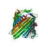
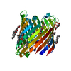
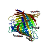
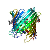
| Title | Crystal structure of the outer membrane protein OprD from Pseudomonas aeruginosa | ||||||
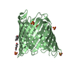
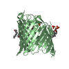
 Components Components | Porin D | ||||||
 Keywords Keywords | MEMBRANE PROTEIN / outer membrane protein / beta-barrel / amino acid transport | ||||||
| Function / homology |  Function and homology information Function and homology informationbasic amino acid transport / outer membrane / porin activity / pore complex / Hydrolases; Acting on peptide bonds (peptidases); Serine endopeptidases / monoatomic ion transport / serine-type peptidase activity / cell outer membrane / proteolysis Similarity search - Function | ||||||
| Biological species |   Pseudomonas aeruginosa (bacteria) Pseudomonas aeruginosa (bacteria) | ||||||
| Method |  X-RAY DIFFRACTION / SYNCHROTRON / SAD / Resolution: 2.9 Å X-RAY DIFFRACTION / SYNCHROTRON / SAD / Resolution: 2.9 Å | ||||||
 Authors Authors | Biswas, S. / van den Berg, B. | ||||||
 Citation Citation | Journal: Nat.Struct.Mol.Biol. / Year: 2007 Title: Structural insight into OprD substrate specificity. Authors: Biswas, S. / Mohammad, M.M. / Patel, D.R. / Movileanu, L. / van den Berg, B. | ||||||
| History |
|
- Structure visualization
| Structure viewer | Molecule: MolmilJmol/JSmol |
|---|
- Downloads & links
-Download
| PDBx/mmCIF format | 2odj.cif.gz | 165.7 KB | Display | PDBx/mmCIF format |
|---|---|---|---|---|
| PDB format | pdb2odj.ent.gz | 132 KB | Display | PDB format |
| PDBx/mmJSON format | 2odj.json.gz | Tree view | PDBx/mmJSON format | |
| Others |  Other downloads Other downloads |
-Validation report
| Arichive directory | https://data.pdbj.org/pub/pdb/validation_reports/od/2odjftp://data.pdbj.org/pub/pdb/validation_reports/od/2odj | HTTPS FTP |
|---|
-Related structure data
| Similar structure data |
|---|


-Links
 PDBj
PDBj- Assembly
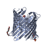
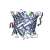
| Deposited unit | 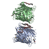
| ||||||||
|---|---|---|---|---|---|---|---|---|---|
| 1 | 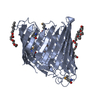
| ||||||||
| 2 | 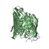
| ||||||||
| Unit cell |
|
-Components
| #1: Protein | Mass: 47443.875 Da / Num. of mol.: 2 Source method: isolated from a genetically manipulated source Source: (gene. exp.) Pseudomonas aeruginosa (bacteria) / Strain: PAO1 / Gene: oprDPlasmid details: Oprd gene with Serratia marcescens ShlB signal sequence cloned into pB22 with C-terminal His8 tag. Protein expression under control of the arabinose promoter Plasmid: pB22 / Production host: References: UniProt: P32722, Hydrolases; Acting on peptide bonds (peptidases); Serine endopeptidases #2: Chemical | ChemComp-C8E / (   Mass: 306.438 Da / Num. of mol.: 8 / Source method: obtained synthetically / Formula: C16H34O5 / Comment: C8E, detergent*YM Mass: 306.438 Da / Num. of mol.: 8 / Source method: obtained synthetically / Formula: C16H34O5 / Comment: C8E, detergent*YMHas protein modification | Y | |
|---|
-Experimental details
-Experiment
| Experiment | Method: X-RAY DIFFRACTION / Number of used crystals: 1 |
|---|
- Sample preparation
| Crystal | Density Matthews: 2.38 Å3/Da / Density % sol: 48.36 % |
|---|---|
| Crystal grow | Temperature: 295 K / Method: vapor diffusion / pH: 4 Details: 0.1 M LiSO4, 0.1 M NaCl, 0.1 M Na citrate, 28-32% PEG 400 (pH 3.5), pH 4.0, VAPOR DIFFUSION, temperature 295K |
-Data collection
| Diffraction | Mean temperature: 100 K |
|---|---|
| Diffraction source | Source: SYNCHROTRON / Site: NSLS  / Beamline: X25 / Wavelength: 0.97922 Å / Beamline: X25 / Wavelength: 0.97922 Å |
| Detector | Type: ADSC QUANTUM 315 / Detector: CCD / Date: Jul 18, 2006 / Details: Platinum coated mirror |
| Radiation | Monochromator: Si 111 double crystal / Protocol: SINGLE WAVELENGTH / Monochromatic (M) / Laue (L): M / Scattering type: x-ray |
| Radiation wavelength | Wavelength: 0.97922 Å / Relative weight: 1 |
| Reflection | Resolution: 2.8→50 Å / Num. obs: 22385 / % possible obs: 98.2 % / Observed criterion σ(F): 0 / Observed criterion σ(I): 0 / Redundancy: 7 % / Rmerge(I) obs: 0.143 / Net I/σ(I): 7 |
| Reflection shell | Resolution: 2.8→2.9 Å / Redundancy: 5.9 % / Rmerge(I) obs: 0.736 / Mean I/σ(I) obs: 2.8 / Num. unique all: 1872 / % possible all: 82.5 |
- Processing
| Software |
| |||||||||||||||||||||||||
|---|---|---|---|---|---|---|---|---|---|---|---|---|---|---|---|---|---|---|---|---|---|---|---|---|---|---|
| Refinement | Method to determine structure: SAD / Resolution: 2.9→10 Å / Cross valid method: THROUGHOUT / σ(F): 0 / σ(I): 0 / Stereochemistry target values: Engh & Huber
| |||||||||||||||||||||||||
| Displacement parameters | Biso mean: 42.4 Å2 | |||||||||||||||||||||||||
| Refine analyze |
| |||||||||||||||||||||||||
| Refinement step | Cycle: LAST / Resolution: 2.9→10 Å
| |||||||||||||||||||||||||
| Refine LS restraints |
| |||||||||||||||||||||||||
| LS refinement shell | Resolution: 2.9→3.03 Å / Rfactor Rfree error: 0.032
|
