 Movie
Movie Controller

[English] 日本語
 Yorodumi
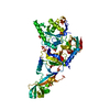
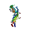
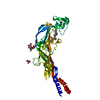
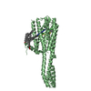
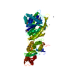
- PDB-3siu: Structure of a hPrp31-15.5K-U4atac 5' stem loop complex, monomeri... -
+ Open data

- Basic information
| Entry | Database: PDB / ID: 3siu | ||||||
|---|---|---|---|---|---|---|---|
| Title | Structure of a hPrp31-15.5K-U4atac 5' stem loop complex, monomeric form | ||||||
 Components Components |
| ||||||
 Keywords Keywords | SPLICING/RNA / Major and minor spliceosome / RNA-protein complex / U4 snRNP and U4atac snRNP / RNA-binding protein / pre-mRNA splicing / U4 snRNA / nucleus / SPLICING-RNA complex | ||||||
| Function / homology |  Function and homology information Function and homology informationribonucleoprotein complex localization / U4atac snRNP / U4atac snRNA binding / box C/D sno(s)RNA binding / dense fibrillar component / box C/D methylation guide snoRNP complex / spliceosomal tri-snRNP complex / snRNP binding / U2-type precatalytic spliceosome / U2-type spliceosomal complex ...ribonucleoprotein complex localization / U4atac snRNP / U4atac snRNA binding / box C/D sno(s)RNA binding / dense fibrillar component / box C/D methylation guide snoRNP complex / spliceosomal tri-snRNP complex / snRNP binding / U2-type precatalytic spliceosome / U2-type spliceosomal complex / U4 snRNA binding / box C/D snoRNP assembly / U4 snRNP / U3 snoRNA binding / precatalytic spliceosome / rRNA modification in the nucleus and cytosol / spliceosomal tri-snRNP complex assembly / MLL1 complex / single fertilization / ribonucleoprotein complex binding / Cajal body / U4/U6 x U5 tri-snRNP complex / Major pathway of rRNA processing in the nucleolus and cytosol / mRNA Splicing - Major Pathway / spliceosomal complex / maturation of SSU-rRNA / small-subunit processome / mRNA splicing, via spliceosome / ATPase binding / ribosomal small subunit biogenesis / protein-macromolecule adaptor activity / nuclear speck / nucleolus / protein-containing complex / RNA binding / nucleoplasm / identical protein binding / nucleus Similarity search - Function | ||||||
| Biological species |  Homo sapiens (human) Homo sapiens (human) | ||||||
| Method |  X-RAY DIFFRACTION / SYNCHROTRON / MOLECULAR REPLACEMENT / Resolution: 2.626 Å X-RAY DIFFRACTION / SYNCHROTRON / MOLECULAR REPLACEMENT / Resolution: 2.626 Å | ||||||
 Authors Authors | Liu, S. / Ghalei, H. / Luhrmann, R. / Wahl, M.C. | ||||||
 Citation Citation | Journal: Rna / Year: 2011 Title: Structural basis for the dual U4 and U4atac snRNA-binding specificity of spliceosomal protein hPrp31. Authors: Liu, S. / Ghalei, H. / Luhrmann, R. / Wahl, M.C. | ||||||
| History |
|
- Structure visualization
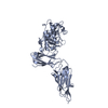
| Structure viewer | Molecule: MolmilJmol/JSmol |
|---|
- Downloads & links
-Download
| PDBx/mmCIF format | 3siu.cif.gz | 185.8 KB | Display | PDBx/mmCIF format |
|---|---|---|---|---|
| PDB format | pdb3siu.ent.gz | 143.5 KB | Display | PDB format |
| PDBx/mmJSON format | 3siu.json.gz | Tree view | PDBx/mmJSON format | |
| Others |  Other downloads Other downloads |
-Validation report
| Arichive directory | https://data.pdbj.org/pub/pdb/validation_reports/si/3siuftp://data.pdbj.org/pub/pdb/validation_reports/si/3siu | HTTPS FTP |
|---|
-Related structure data
| Related structure data |  3sivC  2ozbS S: Starting model for refinement C: citing same article ( |
|---|---|
| Similar structure data |


-Links
 PDBj
PDBj


- Assembly
| Deposited unit | 
| ||||||||
|---|---|---|---|---|---|---|---|---|---|
| 1 | 
| ||||||||
| 2 | 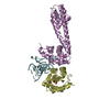
| ||||||||
| Unit cell |
|
-Components
| #1: Protein | Mass: 14335.656 Da / Num. of mol.: 2 Source method: isolated from a genetically manipulated source Source: (gene. exp.) Homo sapiens (human) / Gene: NHP2L1 / Production host:  #2: Protein | Mass: 28268.225 Da / Num. of mol.: 2 / Fragment: UNP residues 85-333 Source method: isolated from a genetically manipulated source Source: (gene. exp.) Homo sapiens (human) / Gene: PRP31, PRPF31 / Production host: #3: RNA chain | Mass: 9073.462 Da / Num. of mol.: 2 / Fragment: GB bases 28-55 / Source method: obtained synthetically / Source: (synth.) Homo sapiens (human) / References: GenBank: NR_023343.1#4: Chemical |   Mass: 96.063 Da / Num. of mol.: 2 / Source method: obtained synthetically / Formula: SO4 Mass: 96.063 Da / Num. of mol.: 2 / Source method: obtained synthetically / Formula: SO4#5: Water | ChemComp-HOH / |  Mass: 18.015 Da / Num. of mol.: 57 / Source method: isolated from a natural source / Formula: H2O Mass: 18.015 Da / Num. of mol.: 57 / Source method: isolated from a natural source / Formula: H2O |
|---|
-Experimental details
-Experiment
| Experiment | Method: X-RAY DIFFRACTION / Number of used crystals: 1 |
|---|
- Sample preparation
| Crystal | Density Matthews: 2.89 Å3/Da / Density % sol: 57.46 % |
|---|---|
| Crystal grow | Temperature: 293 K / Method: vapor diffusion, sitting drop / pH: 7 Details: 0.2 M lithium sulfate, 0.1 M Bis-Tris, pH 7.0, 20% w/v PEG3350, VAPOR DIFFUSION, SITTING DROP, temperature 393K |
-Data collection
| Diffraction | Mean temperature: 100 K |
|---|---|
| Diffraction source | Source: SYNCHROTRON / Site: SLS  / Beamline: X10SA / Wavelength: 0.98 / Beamline: X10SA / Wavelength: 0.98 |
| Detector | Type: MARMOSAIC 225 mm CCD / Detector: CCD / Date: Jan 10, 2007 / Details: mirrors |
| Radiation | Monochromator: Si 111 CHANNEL / Protocol: SINGLE WAVELENGTH / Monochromatic (M) / Laue (L): M / Scattering type: x-ray |
| Radiation wavelength | Wavelength: 0.98 Å / Relative weight: 1 |
| Reflection | Resolution: 2.6→30 Å / Num. all: 34765 / Num. obs: 34765 / % possible obs: 100 % / Observed criterion σ(F): 0 / Observed criterion σ(I): 0 / Redundancy: 5.7 % / Biso Wilson estimate: 66.9 Å2 / Rmerge(I) obs: 0.055 / Rsym value: 0.055 / Net I/σ(I): 19.5 |
| Reflection shell | Resolution: 2.6→2.7 Å / Redundancy: 4.9 % / Rmerge(I) obs: 0.652 / Mean I/σ(I) obs: 1.5 / Num. unique all: 3849 / Rsym value: 0.652 / % possible all: 99.7 |
- Processing
| Software |
| ||||||||||||||||||||||||||||||||||||||||||||||||||||||||||||||||||||||||||||||||||||||||||||||||||
|---|---|---|---|---|---|---|---|---|---|---|---|---|---|---|---|---|---|---|---|---|---|---|---|---|---|---|---|---|---|---|---|---|---|---|---|---|---|---|---|---|---|---|---|---|---|---|---|---|---|---|---|---|---|---|---|---|---|---|---|---|---|---|---|---|---|---|---|---|---|---|---|---|---|---|---|---|---|---|---|---|---|---|---|---|---|---|---|---|---|---|---|---|---|---|---|---|---|---|---|
| Refinement | Method to determine structure: MOLECULAR REPLACEMENT Starting model: PDB ID 2OZB Resolution: 2.626→29.45 Å / SU ML: 0.41 / Isotropic thermal model: ISOTROPIC / Cross valid method: THROUGHOUT / σ(F): 1.35 / σ(I): 0 / Phase error: 30.73 / Stereochemistry target values: ML
| ||||||||||||||||||||||||||||||||||||||||||||||||||||||||||||||||||||||||||||||||||||||||||||||||||
| Solvent computation | Shrinkage radii: 0.9 Å / VDW probe radii: 1.11 Å / Solvent model: FLAT BULK SOLVENT MODEL / Bsol: 63.641 Å2 / ksol: 0.329 e/Å3 | ||||||||||||||||||||||||||||||||||||||||||||||||||||||||||||||||||||||||||||||||||||||||||||||||||
| Displacement parameters | Biso mean: 80.4 Å2
| ||||||||||||||||||||||||||||||||||||||||||||||||||||||||||||||||||||||||||||||||||||||||||||||||||
| Refinement step | Cycle: LAST / Resolution: 2.626→29.45 Å
| ||||||||||||||||||||||||||||||||||||||||||||||||||||||||||||||||||||||||||||||||||||||||||||||||||
| Refine LS restraints |
| ||||||||||||||||||||||||||||||||||||||||||||||||||||||||||||||||||||||||||||||||||||||||||||||||||
| LS refinement shell |
|
