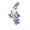
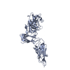
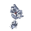
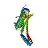
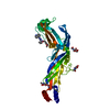
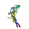
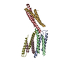
Journal: Nat Commun / Year: 2016 Title: In vivo epidermal migration requires focal adhesion targeting of ACF7. Authors: Jiping Yue / Yao Zhang / Wenguang G Liang / Xuewen Gou / Philbert Lee / Han Liu / Wanqing Lyu / Wei-Jen Tang / Shao-Yu Chen / Feng Yang / Hong Liang / Xiaoyang Wu / Abstract: Turnover of focal adhesions allows cell retraction, which is essential for cell migration. The mammalian spectraplakin protein, ACF7 (Actin-Crosslinking Factor 7), promotes focal adhesion dynamics by ...Turnover of focal adhesions allows cell retraction, which is essential for cell migration. The mammalian spectraplakin protein, ACF7 (Actin-Crosslinking Factor 7), promotes focal adhesion dynamics by targeting of microtubule plus ends towards focal adhesions. However, it remains unclear how the activity of ACF7 is regulated spatiotemporally to achieve focal adhesion-specific guidance of microtubule. To explore the potential mechanisms, we resolve the crystal structure of ACF7's NT (amino-terminal) domain, which mediates F-actin interactions. Structural analysis leads to identification of a key tyrosine residue at the calponin homology (CH) domain of ACF7, whose phosphorylation by Src/FAK (focal adhesion kinase) complex is essential for F-actin binding of ACF7. Using skin epidermis as a model system, we further demonstrate that the phosphorylation of ACF7 plays an indispensable role in focal adhesion dynamics and epidermal migration in vitro and in vivo. Together, our findings provide critical insights into the molecular mechanisms underlying coordinated cytoskeletal dynamics during cell movement.
In the structure databanks used in Yorodumi, some data are registered as the other names, "COVID-19 virus" and "2019-nCoV". Here are the details of the virus and the list of structure data.
Jan 31, 2019. EMDB accession codes are about to change! (news from PDBe EMDB page)
EMDB accession codes are about to change! (news from PDBe EMDB page)
The allocation of 4 digits for EMDB accession codes will soon come to an end. Whilst these codes will remain in use, new EMDB accession codes will include an additional digit and will expand incrementally as the available range of codes is exhausted. The current 4-digit format prefixed with “EMD-” (i.e. EMD-XXXX) will advance to a 5-digit format (i.e. EMD-XXXXX), and so on. It is currently estimated that the 4-digit codes will be depleted around Spring 2019, at which point the 5-digit format will come into force.
The EM Navigator/Yorodumi systems omit the EMD- prefix.
Related info.:Q: What is EMD? / ID/Accession-code notation in Yorodumi/EM Navigator
Yorodumi is a browser for structure data from EMDB, PDB, SASBDB, etc.
This page is also the successor to EM Navigator detail page, and also detail information page/front-end page for Omokage search.
The word "yorodu" (or yorozu) is an old Japanese word meaning "ten thousand". "mi" (miru) is to see.
Related info.:EMDB / PDB / SASBDB / Comparison of 3 databanks / Yorodumi Search / Aug 31, 2016. New EM Navigator & Yorodumi / Yorodumi Papers / Jmol/JSmol / Function and homology information / Changes in new EM Navigator and Yorodumi
 Movie
Movie Controller


 Open data
 Basic information
 Components
 Keywords
 Function and homology information
 Homo sapiens (human)
 X-RAY DIFFRACTION /
  Authors
 Citation

 Structure visualization
 Downloads & links
 Other downloads
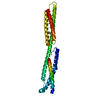


 PDBj
PDBj
 Assembly


 Mass: 94.971 Da / Num. of mol.: 2 / Source method: obtained synthetically / Formula: PO4
 Mass: 18.015 Da / Num. of mol.: 24 / Source method: isolated from a natural source / Formula: H2O
 Sample preparation
 Processing