 Movie
Movie Controller

[English] 日本語
 Yorodumi
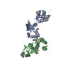
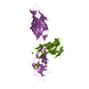
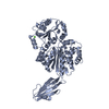
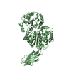
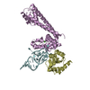
- PDB-3f7p: Crystal structure of a complex between integrin beta4 and plectin -
+ Open data

- Basic information
| Entry | Database: PDB / ID: 3f7p | ||||||
|---|---|---|---|---|---|---|---|
| Title | Crystal structure of a complex between integrin beta4 and plectin | ||||||
 Components Components |
| ||||||
 Keywords Keywords | STRUCTURAL PROTEIN/CELL ADHESION / INTEGRIN / PLAKIN / HEMIDESMOSOME / CELL ADHESION / EPIDERMOLYSIS BULLOSA / Actin-binding / Alternative splicing / Coiled coil / Cytoplasm / Cytoskeleton / Disease mutation / Phosphoprotein / Structural protein / Glycoprotein / Membrane / Polymorphism / Receptor / Transmembrane / STRUCTURAL PROTEIN-CELL ADHESION COMPLEX | ||||||
| Function / homology |  Function and homology information Function and homology informationactomyosin contractile ring assembly actin filament organization / protein-containing complex organization / Type I hemidesmosome assembly / tight junction organization / skeletal myofibril assembly / nail development / hemidesmosome assembly / hemidesmosome / leukocyte migration involved in immune response / peripheral nervous system myelin formation ...actomyosin contractile ring assembly actin filament organization / protein-containing complex organization / Type I hemidesmosome assembly / tight junction organization / skeletal myofibril assembly / nail development / hemidesmosome assembly / hemidesmosome / leukocyte migration involved in immune response / peripheral nervous system myelin formation / trophoblast cell migration / intermediate filament organization / skin morphogenesis / intermediate filament cytoskeleton organization / dystroglycan binding / Laminin interactions / fibroblast migration / cellular response to hydrostatic pressure / costamere / filopodium assembly / T cell chemotaxis / adherens junction organization / peripheral nervous system myelin maintenance / cellular response to fluid shear stress / regulation of vascular permeability / intermediate filament cytoskeleton / myoblast differentiation / mesodermal cell differentiation / Differentiation of Keratinocytes in Interfollicular Epidermis in Mammalian Skin / podosome / Assembly of collagen fibrils and other multimeric structures / integrin complex / cell adhesion mediated by integrin / cardiac muscle cell development / structural constituent of muscle / ankyrin binding / Syndecan interactions / sarcomere organization / nucleus organization / keratinocyte development / response to food / cell leading edge / establishment of skin barrier / transmission of nerve impulse / basement membrane / brush border / sarcoplasm / Caspase-mediated cleavage of cytoskeletal proteins / skeletal muscle fiber development / basal plasma membrane / cell-matrix adhesion / integrin-mediated signaling pathway / respiratory electron transport chain / mitochondrion organization / wound healing / cell motility / cellular response to mechanical stimulus / cell-cell adhesion / sarcolemma / response to wounding / structural constituent of cytoskeleton / G protein-coupled receptor binding / integrin binding / autophagy / multicellular organism growth / cell morphogenesis / Z disc / actin filament binding / cell junction / intracellular protein localization / myelin sheath / cell migration / nuclear membrane / gene expression / mitochondrial outer membrane / cell adhesion / signaling receptor complex / cadherin binding / axon / focal adhesion / dendrite / nucleolus / perinuclear region of cytoplasm / cell surface / RNA binding / extracellular exosome / metal ion binding / identical protein binding / plasma membrane / cytoplasm / cytosol Similarity search - Function | ||||||
| Biological species |  Homo sapiens (human) Homo sapiens (human) | ||||||
| Method |  X-RAY DIFFRACTION / SYNCHROTRON / MOLECULAR REPLACEMENT / Resolution: 2.75 Å X-RAY DIFFRACTION / SYNCHROTRON / MOLECULAR REPLACEMENT / Resolution: 2.75 Å | ||||||
 Authors Authors | de Pereda, J.M. | ||||||
 Citation Citation | Journal: Embo J. / Year: 2009 Title: Structural basis of the interaction between integrin alpha6beta4 and plectin at the hemidesmosomes Authors: de Pereda, J.M. / Lillo, M.P. / Sonnenberg, A. | ||||||
| History |
|
- Structure visualization
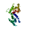
| Structure viewer | Molecule: MolmilJmol/JSmol |
|---|
- Downloads & links
-Download
| PDBx/mmCIF format | 3f7p.cif.gz | 451.5 KB | Display | PDBx/mmCIF format |
|---|---|---|---|---|
| PDB format | pdb3f7p.ent.gz | 369.5 KB | Display | PDB format |
| PDBx/mmJSON format | 3f7p.json.gz | Tree view | PDBx/mmJSON format | |
| Others |  Other downloads Other downloads |
-Validation report
| Arichive directory | https://data.pdbj.org/pub/pdb/validation_reports/f7/3f7pftp://data.pdbj.org/pub/pdb/validation_reports/f7/3f7p | HTTPS FTP |
|---|
-Related structure data
| Related structure data |  3f7qC  3f7rC 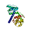 1mb8S  1qg3S S: Starting model for refinement C: citing same article ( |
|---|---|
| Similar structure data |

-Links
 PDBj
PDBj


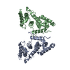
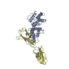
- Assembly
| Deposited unit | 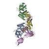
| ||||||||||||||||||||||||||||||||||||||||||||||||||||||||||||||||||||||||||||||||||||||||||||||||||||||||||||||||
|---|---|---|---|---|---|---|---|---|---|---|---|---|---|---|---|---|---|---|---|---|---|---|---|---|---|---|---|---|---|---|---|---|---|---|---|---|---|---|---|---|---|---|---|---|---|---|---|---|---|---|---|---|---|---|---|---|---|---|---|---|---|---|---|---|---|---|---|---|---|---|---|---|---|---|---|---|---|---|---|---|---|---|---|---|---|---|---|---|---|---|---|---|---|---|---|---|---|---|---|---|---|---|---|---|---|---|---|---|---|---|---|---|---|
| 1 | 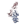
| ||||||||||||||||||||||||||||||||||||||||||||||||||||||||||||||||||||||||||||||||||||||||||||||||||||||||||||||||
| 2 | 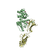
| ||||||||||||||||||||||||||||||||||||||||||||||||||||||||||||||||||||||||||||||||||||||||||||||||||||||||||||||||
| 3 | 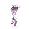
| ||||||||||||||||||||||||||||||||||||||||||||||||||||||||||||||||||||||||||||||||||||||||||||||||||||||||||||||||
| Unit cell |
| ||||||||||||||||||||||||||||||||||||||||||||||||||||||||||||||||||||||||||||||||||||||||||||||||||||||||||||||||
| Components on special symmetry positions |
| ||||||||||||||||||||||||||||||||||||||||||||||||||||||||||||||||||||||||||||||||||||||||||||||||||||||||||||||||
| Noncrystallographic symmetry (NCS) | NCS domain:
NCS domain segments:
|

