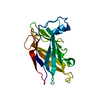
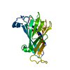
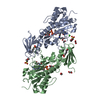
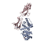
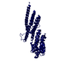
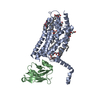
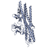
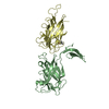
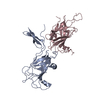
- PDB-6ndx: Lysinoalanine cross-linked FlgE dimer from Treponema denticola -
+
Open data
ID or keywords:
Loading...
-
Basic information
Entry
Database: PDB / ID: 6ndx
Title
Lysinoalanine cross-linked FlgE dimer from Treponema denticola
Components
(Flagellar hook protein FlgE) x 2
Keywords
MOTOR PROTEIN / hook / lysinoalanine / crosslinking / spirochetes / periodontal disease / FlgE / dehydroalanine
Function / homology
Function and homology information
bacterial-type flagellum hook / bacterial-type flagellum basal body / bacterial-type flagellum-dependent swarming motility / cytosol Similarity search - Function
Flagellar hook protein FlgE superfamily / Flagellar hook protein FlgE / Flagellar basal body protein FlaE D2 domain / Flagellar hook-basal body protein, FlgE/F/G / Flagellar hook-basal body protein, FlgE/F/G-like / : / Flagellar hook protein FlgE/F/G D1 domain / Flagellar basal body rod protein, conserved site / Flagella basal body rod proteins signature. / Flagellar basal body rod protein, N-terminal ...Flagellar hook protein FlgE superfamily / Flagellar hook protein FlgE / Flagellar basal body protein FlaE D2 domain / Flagellar hook-basal body protein, FlgE/F/G / Flagellar hook-basal body protein, FlgE/F/G-like / : / Flagellar hook protein FlgE/F/G D1 domain / Flagellar basal body rod protein, conserved site / Flagella basal body rod proteins signature. / Flagellar basal body rod protein, N-terminal / Flagellar basal-body/hook protein, C-terminal domain / Flagella basal body rod protein / Flagellar basal body rod FlgEFG protein C-terminal Similarity search - Domain/homology
Mass: 35533.832 Da / Num. of mol.: 2 / Mutation: C178A Source method: isolated from a genetically manipulated source Details: FlgE(N91-S423) degraded prior to crystallization, N and C termini are missing from structure Source: (gene. exp.) Treponema denticola (bacteria) / Gene: flgE / Production host: Escherichia coli BL21(DE3) (bacteria) / References: UniProt: Q9RQB6
#2: Protein
FlagellarhookproteinFlgE
Mass: 18957.521 Da / Num. of mol.: 2 Source method: isolated from a genetically manipulated source Details: 4 residues on C-termini missing from density, D-alanine present instead of cysteine Source: (gene. exp.) Treponema denticola (bacteria) / Gene: flgE / Production host: Escherichia coli BL21(DE3) (bacteria) / References: UniProt: Q9RQB6
Mass: 18.015 Da / Num. of mol.: 5 / Source method: isolated from a natural source / Formula: H2O
Has protein modification
Y
-
Experimental details
-
Experiment
Experiment
Method: X-RAY DIFFRACTION / Number of used crystals: 1
-
Sample preparation
Crystal
Density Matthews: 2.9 Å3/Da / Density % sol: 57.53 %
Crystal grow
Temperature: 298 K / Method: vapor diffusion, sitting drop Details: crystallized in two conditions: (1) 100 mM Tris pH 8.5, 20% (w/v) PEG8000, 200 mM magnesium chloride and (2) 200 mM magnesium acetate, 20% PEG3350 (w/v)
-
Data collection
Diffraction
Mean temperature: 77 K / Serial crystal experiment: N
In the structure databanks used in Yorodumi, some data are registered as the other names, "COVID-19 virus" and "2019-nCoV". Here are the details of the virus and the list of structure data.
Jan 31, 2019. EMDB accession codes are about to change! (news from PDBe EMDB page)
EMDB accession codes are about to change! (news from PDBe EMDB page)
The allocation of 4 digits for EMDB accession codes will soon come to an end. Whilst these codes will remain in use, new EMDB accession codes will include an additional digit and will expand incrementally as the available range of codes is exhausted. The current 4-digit format prefixed with “EMD-” (i.e. EMD-XXXX) will advance to a 5-digit format (i.e. EMD-XXXXX), and so on. It is currently estimated that the 4-digit codes will be depleted around Spring 2019, at which point the 5-digit format will come into force.
The EM Navigator/Yorodumi systems omit the EMD- prefix.
Related info.:Q: What is EMD? / ID/Accession-code notation in Yorodumi/EM Navigator
Yorodumi is a browser for structure data from EMDB, PDB, SASBDB, etc.
This page is also the successor to EM Navigator detail page, and also detail information page/front-end page for Omokage search.
The word "yorodu" (or yorozu) is an old Japanese word meaning "ten thousand". "mi" (miru) is to see.
Related info.:EMDB / PDB / SASBDB / Comparison of 3 databanks / Yorodumi Search / Aug 31, 2016. New EM Navigator & Yorodumi / Yorodumi Papers / Jmol/JSmol / Function and homology information / Changes in new EM Navigator and Yorodumi
 Movie
Movie Controller


 Open data
 Basic information
 Components
 Keywords
 Function and homology information
 Treponema denticola (bacteria)
 X-RAY DIFFRACTION /
  Authors
 United States, 1items
  Citation
 Structure visualization
 Downloads & links
 Other downloads
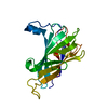

 PDBj
PDBj
 Assembly
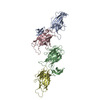
 Mass: 18.015 Da / Num. of mol.: 5 / Source method: isolated from a natural source / Formula: H2O
 Sample preparation
 Processing