 Movie
Movie Controller

[English] 日本語
 Yorodumi
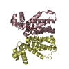
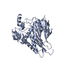
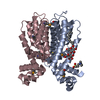
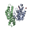
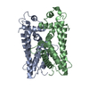
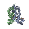
- PDB-3sb9: Cu-mediated Dimer of T4 Lysozyme R76H/R80H by Synthetic Symmetrization -
+ Open data

- Basic information
| Entry | Database: PDB / ID: 3sb9 | ||||||
|---|---|---|---|---|---|---|---|
| Title | Cu-mediated Dimer of T4 Lysozyme R76H/R80H by Synthetic Symmetrization | ||||||
 Components Components | Lysozyme | ||||||
 Keywords Keywords | HYDROLASE / metal-mediated synthetic symmetrization / synthetic symmetrization | ||||||
| Function / homology |  Function and homology information Function and homology informationviral release from host cell by cytolysis / peptidoglycan catabolic process / cell wall macromolecule catabolic process / lysozyme / lysozyme activity / host cell cytoplasm / defense response to bacterium Similarity search - Function | ||||||
| Biological species |  Enterobacteria phage T4 (virus) Enterobacteria phage T4 (virus) | ||||||
| Method |  X-RAY DIFFRACTION / SYNCHROTRON / MOLECULAR REPLACEMENT / molecular replacement / Resolution: 2.45 Å X-RAY DIFFRACTION / SYNCHROTRON / MOLECULAR REPLACEMENT / molecular replacement / Resolution: 2.45 Å | ||||||
 Authors Authors | Soriaga, A.B. / Laganowsky, A. / Zhao, M. / Sawaya, M.R. / Cascio, D. / Yeates, T.O. | ||||||
 Citation Citation | Journal: Protein Sci. / Year: 2011 Title: An approach to crystallizing proteins by metal-mediated synthetic symmetrization. Authors: Laganowsky, A. / Zhao, M. / Soriaga, A.B. / Sawaya, M.R. / Cascio, D. / Yeates, T.O. | ||||||
| History |
|
- Structure visualization
| Structure viewer | Molecule: MolmilJmol/JSmol |
|---|
- Downloads & links
-Download
| PDBx/mmCIF format | 3sb9.cif.gz | 147.6 KB | Display | PDBx/mmCIF format |
|---|---|---|---|---|
| PDB format | pdb3sb9.ent.gz | 116.1 KB | Display | PDB format |
| PDBx/mmJSON format | 3sb9.json.gz | Tree view | PDBx/mmJSON format | |
| Others |  Other downloads Other downloads |
-Validation report
| Arichive directory | https://data.pdbj.org/pub/pdb/validation_reports/sb/3sb9ftp://data.pdbj.org/pub/pdb/validation_reports/sb/3sb9 | HTTPS FTP |
|---|
-Related structure data
| Related structure data | 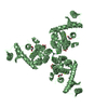 3sb5C  3sb6C 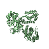 3sb7C 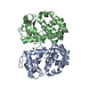 3sb8C 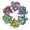 3sbaC 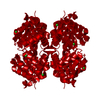 3sbbC 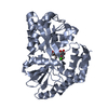 3serC 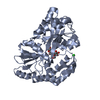 3sesC 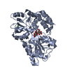 3setC 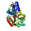 3seuC  3sevC 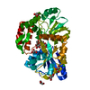 3sewC  3sexC 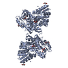 3seyC C: citing same article ( |
|---|---|
| Similar structure data |


-Links
 PDBj
PDBj


- Assembly
| Deposited unit | 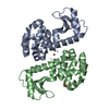
| ||||||||
|---|---|---|---|---|---|---|---|---|---|
| 1 |
| ||||||||
| Unit cell |
|
-Components
| #1: Protein | Mass: 18574.229 Da / Num. of mol.: 2 / Mutation: C54T, R76H, R80H, C97A Source method: isolated from a genetically manipulated source Source: (gene. exp.) Enterobacteria phage T4 (virus) / Gene: E / Plasmid: pET28b / Production host:  #2: Chemical |   Mass: 46.025 Da / Num. of mol.: 2 / Source method: obtained synthetically / Formula: CH2O2 Mass: 46.025 Da / Num. of mol.: 2 / Source method: obtained synthetically / Formula: CH2O2#3: Chemical | ChemComp-CU / |   Mass: 63.546 Da / Num. of mol.: 1 / Source method: obtained synthetically / Formula: Cu Mass: 63.546 Da / Num. of mol.: 1 / Source method: obtained synthetically / Formula: Cu#4: Water | ChemComp-HOH / |  Mass: 18.015 Da / Num. of mol.: 97 / Source method: isolated from a natural source / Formula: H2O Mass: 18.015 Da / Num. of mol.: 97 / Source method: isolated from a natural source / Formula: H2O |
|---|
-Experimental details
-Experiment
| Experiment | Method: X-RAY DIFFRACTION / Number of used crystals: 1 |
|---|
- Sample preparation
| Crystal | Density Matthews: 3.68 Å3/Da / Density % sol: 66.58 % |
|---|---|
| Crystal grow | Temperature: 290 K / Method: vapor diffusion, hanging drop / pH: 7 Details: 3.5M Sodium Formate, pH 7.0, vapor diffusion, hanging drop, temperature 290K |
-Data collection
| Diffraction | Mean temperature: 100 K | |||||||||||||||||||||||||||||||||||||||||||||||||||||||
|---|---|---|---|---|---|---|---|---|---|---|---|---|---|---|---|---|---|---|---|---|---|---|---|---|---|---|---|---|---|---|---|---|---|---|---|---|---|---|---|---|---|---|---|---|---|---|---|---|---|---|---|---|---|---|---|---|
| Diffraction source | Source: SYNCHROTRON / Site: APS  / Beamline: 24-ID-E / Wavelength: 0.9792 Å / Beamline: 24-ID-E / Wavelength: 0.9792 Å | |||||||||||||||||||||||||||||||||||||||||||||||||||||||
| Detector | Type: ADSC QUANTUM 315 / Detector: CCD / Date: Jul 24, 2010 | |||||||||||||||||||||||||||||||||||||||||||||||||||||||
| Radiation | Protocol: SINGLE WAVELENGTH / Monochromatic (M) / Laue (L): M / Scattering type: x-ray | |||||||||||||||||||||||||||||||||||||||||||||||||||||||
| Radiation wavelength | Wavelength: 0.9792 Å / Relative weight: 1 | |||||||||||||||||||||||||||||||||||||||||||||||||||||||
| Reflection | Resolution: 2.45→80 Å / Num. obs: 19881 / % possible obs: 99.9 % / Redundancy: 3.6 % / Rmerge(I) obs: 0.088 / Net I/σ(I): 9.1 | |||||||||||||||||||||||||||||||||||||||||||||||||||||||
| Reflection shell |
|
-Phasing
| Phasing | Method: molecular replacement | |||||||||
|---|---|---|---|---|---|---|---|---|---|---|
| Phasing MR | Model details: Phaser MODE: MR_AUTO
|
- Processing
| Software |
| ||||||||||||||||||||||||||||||||||||||||||||||||||||||||
|---|---|---|---|---|---|---|---|---|---|---|---|---|---|---|---|---|---|---|---|---|---|---|---|---|---|---|---|---|---|---|---|---|---|---|---|---|---|---|---|---|---|---|---|---|---|---|---|---|---|---|---|---|---|---|---|---|---|
| Refinement | Method to determine structure: MOLECULAR REPLACEMENT / Resolution: 2.45→79.114 Å / Occupancy max: 1 / Occupancy min: 0.34 / SU ML: 0.74 / σ(F): 0 / Phase error: 25.3 / Stereochemistry target values: ML
| ||||||||||||||||||||||||||||||||||||||||||||||||||||||||
| Solvent computation | Shrinkage radii: 0.95 Å / VDW probe radii: 1.2 Å / Solvent model: FLAT BULK SOLVENT MODEL / Bsol: 38.893 Å2 / ksol: 0.34 e/Å3 | ||||||||||||||||||||||||||||||||||||||||||||||||||||||||
| Displacement parameters |
| ||||||||||||||||||||||||||||||||||||||||||||||||||||||||
| Refinement step | Cycle: LAST / Resolution: 2.45→79.114 Å
| ||||||||||||||||||||||||||||||||||||||||||||||||||||||||
| Refine LS restraints |
| ||||||||||||||||||||||||||||||||||||||||||||||||||||||||
| LS refinement shell |
| ||||||||||||||||||||||||||||||||||||||||||||||||||||||||
| Refinement TLS params. | Method: refined / Origin x: 18.3847 Å / Origin y: 20.53 Å / Origin z: -0.5173 Å
| ||||||||||||||||||||||||||||||||||||||||||||||||||||||||
| Refinement TLS group | Selection details: ALL |
