 Movie
Movie Controller


+ Open data

- Basic information
| Entry | Database: PDB / ID: 3s96 | ||||||
|---|---|---|---|---|---|---|---|
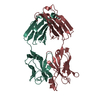
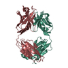
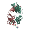
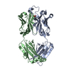
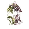
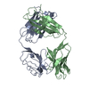
| Title | Crystal structure of 3B5H10 | ||||||
 Components Components |
| ||||||
 Keywords Keywords | IMMUNE SYSTEM / FAB / huntingtin | ||||||
| Function / homology | Immunoglobulins / Immunoglobulin-like / Sandwich / Mainly Beta Function and homology information Function and homology information | ||||||
| Biological species |  | ||||||
| Method |  X-RAY DIFFRACTION / SYNCHROTRON / MOLECULAR REPLACEMENT / Resolution: 1.9 Å X-RAY DIFFRACTION / SYNCHROTRON / MOLECULAR REPLACEMENT / Resolution: 1.9 Å | ||||||
 Authors Authors | Weisgraber, K. / Peters-Libeu, C. / Rutenber, E. / Newhouse, Y. / Finkbeiner, S. | ||||||
 Citation Citation | Journal: J.Mol.Biol. / Year: 2012 Title: Disease-associated polyglutamine stretches in monomeric huntingtin adopt a compact structure. Authors: Peters-Libeu, C. / Miller, J. / Rutenber, E. / Newhouse, Y. / Krishnan, P. / Cheung, K. / Hatters, D. / Brooks, E. / Widjaja, K. / Tran, T. / Mitra, S. / Arrasate, M. / Mosquera, L.A. / ...Authors: Peters-Libeu, C. / Miller, J. / Rutenber, E. / Newhouse, Y. / Krishnan, P. / Cheung, K. / Hatters, D. / Brooks, E. / Widjaja, K. / Tran, T. / Mitra, S. / Arrasate, M. / Mosquera, L.A. / Taylor, D. / Weisgraber, K.H. / Finkbeiner, S. | ||||||
| History |
|
- Structure visualization
| Structure viewer | Molecule: MolmilJmol/JSmol |
|---|
- Downloads & links
-Download
| PDBx/mmCIF format | 3s96.cif.gz | 191.2 KB | Display | PDBx/mmCIF format |
|---|---|---|---|---|
| PDB format | pdb3s96.ent.gz | 152 KB | Display | PDB format |
| PDBx/mmJSON format | 3s96.json.gz | Tree view | PDBx/mmJSON format | |
| Others |  Other downloads Other downloads |
-Validation report
| Arichive directory | https://data.pdbj.org/pub/pdb/validation_reports/s9/3s96ftp://data.pdbj.org/pub/pdb/validation_reports/s9/3s96 | HTTPS FTP |
|---|
-Related structure data


-Links
 PDBj
PDBj

- Assembly
| Deposited unit | 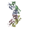
| ||||||||
|---|---|---|---|---|---|---|---|---|---|
| 1 | 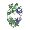
| ||||||||
| 2 | 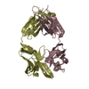
| ||||||||
| Unit cell |
|
-Components
| #1: Antibody | Mass: 23776.730 Da / Num. of mol.: 2 / Source method: isolated from a natural source / Source: (natural) #2: Antibody | Mass: 23558.143 Da / Num. of mol.: 2 / Source method: isolated from a natural source / Source: (natural) #3: Water | ChemComp-HOH / |  Mass: 18.015 Da / Num. of mol.: 890 / Source method: isolated from a natural source / Formula: H2O Mass: 18.015 Da / Num. of mol.: 890 / Source method: isolated from a natural source / Formula: H2OHas protein modification | Y | |
|---|
-Experimental details
-Experiment
| Experiment | Method: X-RAY DIFFRACTION / Number of used crystals: 1 |
|---|
- Sample preparation
| Crystal | Density Matthews: 2.06 Å3/Da / Density % sol: 40 % |
|---|---|
| Crystal grow | pH: 4.5 Details: 7% PEG 3350, 66MM CITRIC ACID, 2 MM TRIS, 5% V/V ETHYL ACETATE, PH 4.50, VAPOR DIFFUSION, HANGING DROP, TEMPERATURE 323K |
-Data collection
| Diffraction | Mean temperature: 100 K |
|---|---|
| Diffraction source | Source: SYNCHROTRON / Site: ALS  / Beamline: 8.3.1 / Wavelength: 1.078 / Beamline: 8.3.1 / Wavelength: 1.078 |
| Detector | Type: ADSC QUANTUM 4 / Detector: CCD / Date: Jan 31, 2004 |
| Radiation | Protocol: SINGLE WAVELENGTH / Monochromatic (M) / Laue (L): M / Scattering type: x-ray |
| Radiation wavelength | Wavelength: 1.078 Å / Relative weight: 1 |
| Reflection | Resolution: 1.9→15 Å / Num. obs: 70733 / % possible obs: 95.6 % / Observed criterion σ(I): 0 / Redundancy: 3.2 % / Rsym value: 0.081 / Net I/σ(I): 10.8 |
| Reflection shell | Resolution: 1.9→1.95 Å / Redundancy: 1.6 % / Mean I/σ(I) obs: 5.2 / Rsym value: 0.236 / % possible all: 79 |
- Processing
| Software |
| ||||||||||||||||||||||||||||||||||||||||||||||||||||||||||||||||||||||||||||||||||||||||||||||||||||||||||||||||||||||||||||||||||||||||||||||||||||||||||||||||||||||||||
|---|---|---|---|---|---|---|---|---|---|---|---|---|---|---|---|---|---|---|---|---|---|---|---|---|---|---|---|---|---|---|---|---|---|---|---|---|---|---|---|---|---|---|---|---|---|---|---|---|---|---|---|---|---|---|---|---|---|---|---|---|---|---|---|---|---|---|---|---|---|---|---|---|---|---|---|---|---|---|---|---|---|---|---|---|---|---|---|---|---|---|---|---|---|---|---|---|---|---|---|---|---|---|---|---|---|---|---|---|---|---|---|---|---|---|---|---|---|---|---|---|---|---|---|---|---|---|---|---|---|---|---|---|---|---|---|---|---|---|---|---|---|---|---|---|---|---|---|---|---|---|---|---|---|---|---|---|---|---|---|---|---|---|---|---|---|---|---|---|---|---|---|
| Refinement | Method to determine structure: MOLECULAR REPLACEMENT Starting model: A STRUCTURE DETERMINED FROM A SINGLE PT DERIVATIVE IN ANOTHER SPACE GROUP WAS USED AS THE PROBE MODEL FOR MOLECULAR REPLACEMENT. Resolution: 1.9→11 Å / Cor.coef. Fo:Fc: 0.942 / Cor.coef. Fo:Fc free: 0.874 / SU B: 4.103 / SU ML: 0.124 / Cross valid method: THROUGHOUT / σ(F): 0 / ESU R Free: 0.187 / Stereochemistry target values: MAXIMUM LIKELIHOOD / Details: HYDROGENS HAVE BEEN ADDED IN THE RIDING POSITIONS
| ||||||||||||||||||||||||||||||||||||||||||||||||||||||||||||||||||||||||||||||||||||||||||||||||||||||||||||||||||||||||||||||||||||||||||||||||||||||||||||||||||||||||||
| Solvent computation | Ion probe radii: 0.8 Å / Shrinkage radii: 0.8 Å / VDW probe radii: 1.2 Å / Solvent model: MASK | ||||||||||||||||||||||||||||||||||||||||||||||||||||||||||||||||||||||||||||||||||||||||||||||||||||||||||||||||||||||||||||||||||||||||||||||||||||||||||||||||||||||||||
| Displacement parameters | Biso mean: 17.23 Å2
| ||||||||||||||||||||||||||||||||||||||||||||||||||||||||||||||||||||||||||||||||||||||||||||||||||||||||||||||||||||||||||||||||||||||||||||||||||||||||||||||||||||||||||
| Refinement step | Cycle: LAST / Resolution: 1.9→11 Å
| ||||||||||||||||||||||||||||||||||||||||||||||||||||||||||||||||||||||||||||||||||||||||||||||||||||||||||||||||||||||||||||||||||||||||||||||||||||||||||||||||||||||||||
| Refine LS restraints |
| ||||||||||||||||||||||||||||||||||||||||||||||||||||||||||||||||||||||||||||||||||||||||||||||||||||||||||||||||||||||||||||||||||||||||||||||||||||||||||||||||||||||||||
| LS refinement shell | Resolution: 1.9→1.95 Å / Total num. of bins used: 20
|
