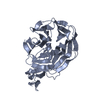
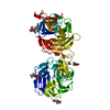
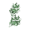
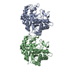
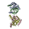
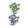
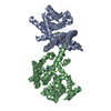
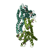
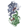
Low-density lipoprotein receptor-related protein 6
Keywords
SIGNALING PROTEIN / Wnt / receptor / LRP5 / LRP6 / LDL receptor-like protein / Dickkopf (Dkk) / YWTD b-propeller
Function / homology
Function and homology information
Wnt-Frizzled-LRP5/6 complex / Negative regulation of TCF-dependent signaling by WNT ligand antagonists / Signaling by RNF43 mutants / neural crest formation / kinase inhibitor activity / Wnt receptor activity / low-density lipoprotein particle receptor activity / toxin transmembrane transporter activity / Wnt-protein binding / cellular response to cholesterol ...Wnt-Frizzled-LRP5/6 complex / Negative regulation of TCF-dependent signaling by WNT ligand antagonists / Signaling by RNF43 mutants / neural crest formation / kinase inhibitor activity / Wnt receptor activity / low-density lipoprotein particle receptor activity / toxin transmembrane transporter activity / Wnt-protein binding / cellular response to cholesterol / dopaminergic neuron differentiation / midbrain dopaminergic neuron differentiation / frizzled binding / Wnt signalosome / Disassembly of the destruction complex and recruitment of AXIN to the membrane / neural crest cell differentiation / negative regulation of smooth muscle cell apoptotic process / canonical Wnt signaling pathway / protein serine/threonine kinase inhibitor activity / coreceptor activity / positive regulation of cell cycle / Regulation of FZD by ubiquitination / protein localization to plasma membrane / TCF dependent signaling in response to WNT / cell-cell adhesion / response to peptide hormone / Wnt signaling pathway / endocytosis / nervous system development / positive regulation of cytosolic calcium ion concentration / cytoplasmic vesicle / early endosome membrane / chemical synaptic transmission / membrane raft / signaling receptor binding / neuronal cell body / synapse / positive regulation of DNA-templated transcription / cell surface / endoplasmic reticulum / protein homodimerization activity / positive regulation of transcription by RNA polymerase II / extracellular region / identical protein binding / plasma membrane Similarity search - Function
Low density lipoprotein receptor-related protein 5/6 / : / TolB, C-terminal domain / Low-density lipoprotein receptor repeat class B / LDL-receptor class B (LDLRB) repeat profile. / LDLR class B repeat / Low-density lipoprotein-receptor YWTD domain / Low-density lipoprotein receptor domain class A / Low-density lipoprotein (LDL) receptor class A, conserved site / LDL-receptor class A (LDLRA) domain signature. ...Low density lipoprotein receptor-related protein 5/6 / : / TolB, C-terminal domain / Low-density lipoprotein receptor repeat class B / LDL-receptor class B (LDLRB) repeat profile. / LDLR class B repeat / Low-density lipoprotein-receptor YWTD domain / Low-density lipoprotein receptor domain class A / Low-density lipoprotein (LDL) receptor class A, conserved site / LDL-receptor class A (LDLRA) domain signature. / LDL-receptor class A (LDLRA) domain profile. / Low-density lipoprotein receptor domain class A / Low-density lipoprotein (LDL) receptor class A repeat / LDL receptor-like superfamily / Six-bladed beta-propeller, TolB-like / Coagulation Factor Xa inhibitory site / 6 Propeller / Neuraminidase / Epidermal growth factor-like domain. / EGF-like domain signature 2. / EGF-like domain / Mainly Beta Similarity search - Domain/homology
In the structure databanks used in Yorodumi, some data are registered as the other names, "COVID-19 virus" and "2019-nCoV". Here are the details of the virus and the list of structure data.
Jan 31, 2019. EMDB accession codes are about to change! (news from PDBe EMDB page)
EMDB accession codes are about to change! (news from PDBe EMDB page)
The allocation of 4 digits for EMDB accession codes will soon come to an end. Whilst these codes will remain in use, new EMDB accession codes will include an additional digit and will expand incrementally as the available range of codes is exhausted. The current 4-digit format prefixed with “EMD-” (i.e. EMD-XXXX) will advance to a 5-digit format (i.e. EMD-XXXXX), and so on. It is currently estimated that the 4-digit codes will be depleted around Spring 2019, at which point the 5-digit format will come into force.
The EM Navigator/Yorodumi systems omit the EMD- prefix.
Related info.:Q: What is EMD? / ID/Accession-code notation in Yorodumi/EM Navigator
Yorodumi is a browser for structure data from EMDB, PDB, SASBDB, etc.
This page is also the successor to EM Navigator detail page, and also detail information page/front-end page for Omokage search.
The word "yorodu" (or yorozu) is an old Japanese word meaning "ten thousand". "mi" (miru) is to see.
Related info.:EMDB / PDB / SASBDB / Comparison of 3 databanks / Yorodumi Search / Aug 31, 2016. New EM Navigator & Yorodumi / Yorodumi Papers / Jmol/JSmol / Function and homology information / Changes in new EM Navigator and Yorodumi
 Movie
Movie Controller


 Open data
 Basic information
 Components
 Keywords
 Function and homology information
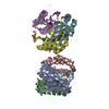
 Homo sapiens (human)
 X-RAY DIFFRACTION /
  Authors
 Citation
 Structure visualization
 Downloads & links
 Other downloads


 PDBj
PDBj


 Assembly
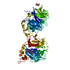

 Spodoptera frugiperda (fall armyworm) / References: UniProt: O75581
 Type: D-saccharide, beta linking / Mass: 221.208 Da / Num. of mol.: 3
 Sample preparation
 / Beamline: 8.2.2
 Processing