 Movie
Movie Controller

[English] 日本語
 Yorodumi
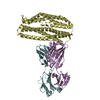
- PDB-3s88: Crystal structure of Sudan Ebolavirus Glycoprotein (strain Gulu) ... -
+ Open data

- Basic information
| Entry | Database: PDB / ID: 3s88 | |||||||||
|---|---|---|---|---|---|---|---|---|---|---|
| Title | Crystal structure of Sudan Ebolavirus Glycoprotein (strain Gulu) bound to 16F6 | |||||||||
 Components Components |
| |||||||||
 Keywords Keywords | IMMUNE SYSTEM/VIRAL PROTEIN / Glycosylation / Viral membrane / IMMUNE SYSTEM-VIRAL PROTEIN complex | |||||||||
| Function / homology |  Function and homology information Function and homology informationsymbiont-mediated-mediated suppression of host tetherin activity / clathrin-dependent endocytosis of virus by host cell / entry receptor-mediated virion attachment to host cell / symbiont-mediated suppression of host innate immune response / fusion of virus membrane with host endosome membrane / viral envelope / host cell plasma membrane / virion membrane / extracellular region Similarity search - Function | |||||||||
| Biological species |   Sudan ebolavirus Sudan ebolavirus | |||||||||
| Method |  X-RAY DIFFRACTION / SYNCHROTRON / MOLECULAR REPLACEMENT / Resolution: 3.351 Å X-RAY DIFFRACTION / SYNCHROTRON / MOLECULAR REPLACEMENT / Resolution: 3.351 Å | |||||||||
 Authors Authors | Saphire, E.O. / Dias, J.M. / Bale, S. | |||||||||
 Citation Citation | Journal: Nat.Struct.Mol.Biol. / Year: 2011 Title: A shared structural solution for neutralizing ebolaviruses. Authors: Dias, J.M. / Kuehne, A.I. / Abelson, D.M. / Bale, S. / Wong, A.C. / Halfmann, P. / Muhammad, M.A. / Fusco, M.L. / Zak, S.E. / Kang, E. / Kawaoka, Y. / Chandran, K. / Dye, J.M. / Saphire, E.O. | |||||||||
| History |
|
- Structure visualization
| Structure viewer | Molecule: MolmilJmol/JSmol |
|---|
- Downloads & links
-Download
| PDBx/mmCIF format | 3s88.cif.gz | 324 KB | Display | PDBx/mmCIF format |
|---|---|---|---|---|
| PDB format | pdb3s88.ent.gz | 264.3 KB | Display | PDB format |
| PDBx/mmJSON format | 3s88.json.gz | Tree view | PDBx/mmJSON format | |
| Others |  Other downloads Other downloads |
-Validation report
| Arichive directory | https://data.pdbj.org/pub/pdb/validation_reports/s8/3s88ftp://data.pdbj.org/pub/pdb/validation_reports/s8/3s88 | HTTPS FTP |
|---|
-Related structure data
| Related structure data | 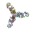 3csyS S: Starting model for refinement |
|---|---|
| Similar structure data |


-Links
 PDBj
PDBj


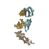
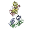
- Assembly
| Deposited unit | 
| ||||||||
|---|---|---|---|---|---|---|---|---|---|
| 1 |
| ||||||||
| 2 | x 12
| ||||||||
| 3 | 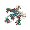
| ||||||||
| 4 | 
| ||||||||
| 5 | 
| ||||||||
| Unit cell |
|
-Components
| #1: Antibody | Mass: 23600.488 Da / Num. of mol.: 1 / Fragment: UNP Residues 32-313 Source method: isolated from a genetically manipulated source Details: Vaccination / Source: (gene. exp.) | ||
|---|---|---|---|
| #2: Protein | Mass: 33447.328 Da / Num. of mol.: 1 / Fragment: UNP Residues 473-639 / Mutation: I631V, Q638V Source method: isolated from a genetically manipulated source Source: (gene. exp.) Sudan ebolavirus / Strain: Gulu / Gene: GP / Plasmid: PDISPLAY / Cell line (production host): HEK293T / Production host:  Homo sapiens (human) / References: UniProt: Q7T9D9 Homo sapiens (human) / References: UniProt: Q7T9D9 | ||
| #3: Protein | Mass: 18709.189 Da / Num. of mol.: 1 Source method: isolated from a genetically manipulated source Source: (gene. exp.) Sudan ebolavirus / Strain: Gulu / Gene: GP / Plasmid: PDISPLAY / Cell line (production host): HEK293T / Production host: Homo sapiens (human) / References: UniProt: Q7T9D9 | ||
| #4: Antibody | Mass: 23457.027 Da / Num. of mol.: 1 Source method: isolated from a genetically manipulated source Details: Vaccination / Source: (gene. exp.) | ||
| #5: Polysaccharide | Source method: isolated from a genetically manipulated source Has protein modification | Y | |
-Experimental details
-Experiment
| Experiment | Method: X-RAY DIFFRACTION / Number of used crystals: 1 |
|---|
- Sample preparation
| Crystal | Density Matthews: 3.05 Å3/Da / Density % sol: 59.63 % |
|---|---|
| Crystal grow | Temperature: 298 K / Method: vapor diffusion, hanging drop / pH: 8.4 Details: 15% PEG 3350, 0.2 M Lithium citrate, pH 8.4, VAPOR DIFFUSION, HANGING DROP, temperature 298K |
-Data collection
| Diffraction | Mean temperature: 77 K |
|---|---|
| Diffraction source | Source: SYNCHROTRON / Site: APS  / Beamline: 19-ID / Wavelength: 0.9793 Å / Beamline: 19-ID / Wavelength: 0.9793 Å |
| Detector | Type: ADSC QUANTUM 315r / Detector: CCD / Date: Apr 1, 2009 |
| Radiation | Protocol: SINGLE WAVELENGTH / Monochromatic (M) / Laue (L): M / Scattering type: x-ray |
| Radiation wavelength | Wavelength: 0.9793 Å / Relative weight: 1 |
| Reflection | Resolution: 3.35→50 Å / Num. all: 17047 / Num. obs: 17047 / % possible obs: 100 % / Observed criterion σ(F): 1 / Observed criterion σ(I): 1 / Redundancy: 5.3 % / Biso Wilson estimate: 68.6 Å2 / Rsym value: 0.088 / Net I/σ(I): 10.1 |
| Reflection shell | Resolution: 3.35→3.43 Å / Redundancy: 5.9 % / Mean I/σ(I) obs: 1.7 / Num. unique all: 1881 / Rsym value: 0.825 / % possible all: 100 |
- Processing
| Software |
| ||||||||||||||||||||||||||||||||||||||||||||||||||||||||||||||||||||||||||||||||||||||||||||||||||||||||||||||||||||||||||||||||||||||||||||||||||||||||||||||||||||||||||
|---|---|---|---|---|---|---|---|---|---|---|---|---|---|---|---|---|---|---|---|---|---|---|---|---|---|---|---|---|---|---|---|---|---|---|---|---|---|---|---|---|---|---|---|---|---|---|---|---|---|---|---|---|---|---|---|---|---|---|---|---|---|---|---|---|---|---|---|---|---|---|---|---|---|---|---|---|---|---|---|---|---|---|---|---|---|---|---|---|---|---|---|---|---|---|---|---|---|---|---|---|---|---|---|---|---|---|---|---|---|---|---|---|---|---|---|---|---|---|---|---|---|---|---|---|---|---|---|---|---|---|---|---|---|---|---|---|---|---|---|---|---|---|---|---|---|---|---|---|---|---|---|---|---|---|---|---|---|---|---|---|---|---|---|---|---|---|---|---|---|---|---|
| Refinement | Method to determine structure: MOLECULAR REPLACEMENT Starting model: PDB ENTRY 3CSY Resolution: 3.351→45.63 Å / Cor.coef. Fo:Fc: 0.926 / Cor.coef. Fo:Fc free: 0.908 / SU B: 70.373 / SU ML: 0.516 / Cross valid method: THROUGHOUT / σ(F): 1 / ESU R Free: 0.579 / Stereochemistry target values: MAXIMUM LIKELIHOOD / Details: HYDROGENS HAVE BEEN ADDED IN THE RIDING POSITIONS
| ||||||||||||||||||||||||||||||||||||||||||||||||||||||||||||||||||||||||||||||||||||||||||||||||||||||||||||||||||||||||||||||||||||||||||||||||||||||||||||||||||||||||||
| Solvent computation | Ion probe radii: 0.8 Å / Shrinkage radii: 0.8 Å / VDW probe radii: 1.4 Å / Solvent model: MASK | ||||||||||||||||||||||||||||||||||||||||||||||||||||||||||||||||||||||||||||||||||||||||||||||||||||||||||||||||||||||||||||||||||||||||||||||||||||||||||||||||||||||||||
| Displacement parameters | Biso mean: 102.838 Å2 | ||||||||||||||||||||||||||||||||||||||||||||||||||||||||||||||||||||||||||||||||||||||||||||||||||||||||||||||||||||||||||||||||||||||||||||||||||||||||||||||||||||||||||
| Refinement step | Cycle: LAST / Resolution: 3.351→45.63 Å
| ||||||||||||||||||||||||||||||||||||||||||||||||||||||||||||||||||||||||||||||||||||||||||||||||||||||||||||||||||||||||||||||||||||||||||||||||||||||||||||||||||||||||||
| Refine LS restraints |
| ||||||||||||||||||||||||||||||||||||||||||||||||||||||||||||||||||||||||||||||||||||||||||||||||||||||||||||||||||||||||||||||||||||||||||||||||||||||||||||||||||||||||||
| LS refinement shell | Resolution: 3.351→3.437 Å / Total num. of bins used: 20
| ||||||||||||||||||||||||||||||||||||||||||||||||||||||||||||||||||||||||||||||||||||||||||||||||||||||||||||||||||||||||||||||||||||||||||||||||||||||||||||||||||||||||||
| Refinement TLS params. | Method: refined / Refine-ID: X-RAY DIFFRACTION
| ||||||||||||||||||||||||||||||||||||||||||||||||||||||||||||||||||||||||||||||||||||||||||||||||||||||||||||||||||||||||||||||||||||||||||||||||||||||||||||||||||||||||||
| Refinement TLS group |
|
