 Movie
Movie Controller

[English] 日本語
 Yorodumi
- PDB-3rt0: Crystal structure of PYL10-HAB1 complex in the absence of abscisi... -
+ Open data

- Basic information
| Entry | Database: PDB / ID: 3rt0 | ||||||
|---|---|---|---|---|---|---|---|
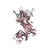
| Title | Crystal structure of PYL10-HAB1 complex in the absence of abscisic acid (ABA) | ||||||
 Components Components |
| ||||||
 Keywords Keywords | HYDROLASE/HYDROLASE INHIBITOR / PYL10-HAB1 binary COMPLEX / apo-PYL10 inhibits HAB1 dephosphorylation activity / HYDROLASE-HYDROLASE INHIBITOR complex | ||||||
| Function / homology |  Function and homology information Function and homology informationabscisic acid binding / abscisic acid-activated signaling pathway / protein phosphatase inhibitor activity / protein-serine/threonine phosphatase / protein serine/threonine phosphatase activity / signaling receptor activity / protein homodimerization activity / metal ion binding / nucleus / plasma membrane / cytoplasm Similarity search - Function | ||||||
| Biological species |  | ||||||
| Method |  X-RAY DIFFRACTION / SYNCHROTRON / MOLECULAR REPLACEMENT / Resolution: 2.113 Å X-RAY DIFFRACTION / SYNCHROTRON / MOLECULAR REPLACEMENT / Resolution: 2.113 Å | ||||||
 Authors Authors | Hao, Q. / Yin, P. / Li, W. / Wang, L. / Yan, C. / Wang, J. / Yan, N. | ||||||
 Citation Citation | Journal: Mol.Cell / Year: 2011 Title: The Molecular Basis of ABA-Independent Inhibition of PP2Cs by a Subclass of PYL Proteins Authors: Hao, Q. / Yin, P. / Li, W. / Wang, L. / Yan, C. / Lin, Z. / Wu, J.Z. / Wang, J. / Yan, S.F. / Yan, N. | ||||||
| History |
|
- Structure visualization
| Structure viewer | Molecule: MolmilJmol/JSmol |
|---|
- Downloads & links
-Download
| PDBx/mmCIF format | 3rt0.cif.gz | 407.5 KB | Display | PDBx/mmCIF format |
|---|---|---|---|---|
| PDB format | pdb3rt0.ent.gz | 332.6 KB | Display | PDB format |
| PDBx/mmJSON format | 3rt0.json.gz | Tree view | PDBx/mmJSON format | |
| Others |  Other downloads Other downloads |
-Validation report
| Summary document | 3rt0_validation.pdf.gz | 464 KB | Display | wwPDB validaton report |
|---|---|---|---|---|
| Full document | 3rt0_full_validation.pdf.gz | 487.8 KB | Display | |
| Data in XML | 3rt0_validation.xml.gz | 43.5 KB | Display | |
| Data in CIF | 3rt0_validation.cif.gz | 61.3 KB | Display | |
| Arichive directory | https://data.pdbj.org/pub/pdb/validation_reports/rt/3rt0ftp://data.pdbj.org/pub/pdb/validation_reports/rt/3rt0 | HTTPS FTP |
-Related structure data
| Related structure data | 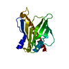 3rt2SC S: Starting model for refinement C: citing same article ( |
|---|---|
| Similar structure data |
-Links
 PDBj
PDBj- Assembly
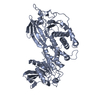
| Deposited unit | 
| ||||||||
|---|---|---|---|---|---|---|---|---|---|
| 1 | 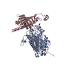
| ||||||||
| 2 | 
| ||||||||
| Unit cell |
|
-Components
| #1: Protein | Mass: 37753.219 Da / Num. of mol.: 2 / Fragment: residues 172- 511 / Mutation: C274S Source method: isolated from a genetically manipulated source Source: (gene. exp.)  References: UniProt: Q9CAJ0, protein-serine/threonine phosphatase #2: Protein | Mass: 20658.545 Da / Num. of mol.: 2 / Mutation: C166S Source method: isolated from a genetically manipulated source Source: (gene. exp.) #3: Chemical |   Mass: 24.305 Da / Num. of mol.: 2 / Source method: obtained synthetically / Formula: Mg Mass: 24.305 Da / Num. of mol.: 2 / Source method: obtained synthetically / Formula: Mg#4: Water | ChemComp-HOH / |  Mass: 18.015 Da / Num. of mol.: 507 / Source method: isolated from a natural source / Formula: H2O Mass: 18.015 Da / Num. of mol.: 507 / Source method: isolated from a natural source / Formula: H2O |
|---|
-Experimental details
-Experiment
| Experiment | Method: X-RAY DIFFRACTION / Number of used crystals: 1 |
|---|
- Sample preparation
| Crystal | Density Matthews: 2.22 Å3/Da / Density % sol: 44.64 % |
|---|---|
| Crystal grow | Temperature: 291 K / Method: vapor diffusion, hanging drop / pH: 8 Details: 100mM Tris, 21% PEG3350, 2% Dioxane , pH 8.0, VAPOR DIFFUSION, HANGING DROP, temperature 291K |
-Data collection
| Diffraction | Mean temperature: 100 K |
|---|---|
| Diffraction source | Source: SYNCHROTRON / Site: SPring-8  / Beamline: BL41XU / Wavelength: 1 Å / Beamline: BL41XU / Wavelength: 1 Å |
| Detector | Type: RAYONIX MX225HE / Detector: CCD / Date: Oct 27, 2010 |
| Radiation | Monochromator: rotated-inclined double-crystal monochromator Protocol: SINGLE WAVELENGTH / Monochromatic (M) / Laue (L): M / Scattering type: x-ray |
| Radiation wavelength | Wavelength: 1 Å / Relative weight: 1 |
| Reflection | Resolution: 2.1→35 Å / Num. obs: 58037 / % possible obs: 99.8 % / Biso Wilson estimate: 25.43 Å2 |
| Reflection shell | Resolution: 2.1→2.18 Å / Redundancy: 3.9 % / Rmerge(I) obs: 0.399 / Mean I/σ(I) obs: 4.16 / Num. unique all: 5772 / Rsym value: 0.399 / % possible all: 99.9 |
- Processing
| Software |
| |||||||||||||||||||||||||||||||||||||||||||||||||||||||||||||||||||||||||||||||||||||||||||||||||||||||||||||||||||||||||||||||||||||||||||||||||||
|---|---|---|---|---|---|---|---|---|---|---|---|---|---|---|---|---|---|---|---|---|---|---|---|---|---|---|---|---|---|---|---|---|---|---|---|---|---|---|---|---|---|---|---|---|---|---|---|---|---|---|---|---|---|---|---|---|---|---|---|---|---|---|---|---|---|---|---|---|---|---|---|---|---|---|---|---|---|---|---|---|---|---|---|---|---|---|---|---|---|---|---|---|---|---|---|---|---|---|---|---|---|---|---|---|---|---|---|---|---|---|---|---|---|---|---|---|---|---|---|---|---|---|---|---|---|---|---|---|---|---|---|---|---|---|---|---|---|---|---|---|---|---|---|---|---|---|---|---|
| Refinement | Method to determine structure: MOLECULAR REPLACEMENT Starting model: 3RT2 Resolution: 2.113→32.499 Å / Occupancy max: 1 / Occupancy min: 0.12 / FOM work R set: 0.8629 / SU ML: 0.24 / σ(F): 0 / Phase error: 21.01 / Stereochemistry target values: ML
| |||||||||||||||||||||||||||||||||||||||||||||||||||||||||||||||||||||||||||||||||||||||||||||||||||||||||||||||||||||||||||||||||||||||||||||||||||
| Solvent computation | Shrinkage radii: 0.83 Å / VDW probe radii: 1.1 Å / Solvent model: FLAT BULK SOLVENT MODEL / Bsol: 41.02 Å2 / ksol: 0.338 e/Å3 | |||||||||||||||||||||||||||||||||||||||||||||||||||||||||||||||||||||||||||||||||||||||||||||||||||||||||||||||||||||||||||||||||||||||||||||||||||
| Displacement parameters | Biso max: 203.27 Å2 / Biso mean: 36.291 Å2 / Biso min: 9.77 Å2
| |||||||||||||||||||||||||||||||||||||||||||||||||||||||||||||||||||||||||||||||||||||||||||||||||||||||||||||||||||||||||||||||||||||||||||||||||||
| Refinement step | Cycle: LAST / Resolution: 2.113→32.499 Å
| |||||||||||||||||||||||||||||||||||||||||||||||||||||||||||||||||||||||||||||||||||||||||||||||||||||||||||||||||||||||||||||||||||||||||||||||||||
| Refine LS restraints |
| |||||||||||||||||||||||||||||||||||||||||||||||||||||||||||||||||||||||||||||||||||||||||||||||||||||||||||||||||||||||||||||||||||||||||||||||||||
| LS refinement shell | Refine-ID: X-RAY DIFFRACTION / Total num. of bins used: 20
| |||||||||||||||||||||||||||||||||||||||||||||||||||||||||||||||||||||||||||||||||||||||||||||||||||||||||||||||||||||||||||||||||||||||||||||||||||
| Refinement TLS params. | Method: refined / Origin x: 37.9109 Å / Origin y: 15.0621 Å / Origin z: 12.3016 Å
| |||||||||||||||||||||||||||||||||||||||||||||||||||||||||||||||||||||||||||||||||||||||||||||||||||||||||||||||||||||||||||||||||||||||||||||||||||
| Refinement TLS group |
|
